. 2024 Apr 17;23(1):106.
doi: 10.1186/s12936-024-04933-y.

Superior protection in a relapsing Plasmodium cynomolgi rhesus macaque model by a chemoprophylaxis with sporozoite immunization regimen with atovaquone-proguanil followed by primaquine

Affiliations

Superior protection in a relapsing Plasmodium cynomolgi rhesus macaque model by a chemoprophylaxis with sporozoite immunization regimen with atovaquone-proguanil followed by primaquine

Kosol Yongvanitchit et al. Malar J. .

Abstract

Background: To gain a deeper understanding of protective immunity against relapsing malaria, this study examined sporozoite-specific T cell responses induced by a chemoprophylaxis with sporozoite (CPS) immunization in a relapsing Plasmodium cynomolgi rhesus macaque model.

Methods: The animals received three CPS immunizations with P. cynomolgi sporozoites, administered by mosquito bite, while under two anti-malarial drug regimens. Group 1 (n = 6) received artesunate/chloroquine (AS/CQ) followed by a radical cure with CQ plus primaquine (PQ). Group 2 (n = 6) received atovaquone-proguanil (AP) followed by PQ. After the final immunization, the animals were challenged with intravenous injection of 104 P. cynomolgi sporozoites, the dose that induced reliable infection and relapse rate. These animals, along with control animals (n = 6), were monitored for primary infection and subsequent relapses. Immunogenicity blood draws were done after each of the three CPS session, before and after the challenge, with liver, spleen and bone marrow sampling and analysis done after the challenge.

Results: Group 2 animals demonstrated superior protection, with two achieving protection and two experiencing partial protection, while only one animal in group 1 had partial protection. These animals displayed high sporozoite-specific IFN-γ T cell responses in the liver, spleen, and bone marrow after the challenge with one protected animal having the highest frequency of IFN-γ+ CD8+, IFN-γ+ CD4+, and IFN-γ+ γδ T cells in the liver. Partially protected animals also demonstrated a relatively high frequency of IFN-γ+ CD8+, IFN-γ+ CD4+, and IFN-γ+ γδ T cells in the liver. It is important to highlight that the second animal in group 2, which experienced protection, exhibited deficient sporozoite-specific T cell responses in the liver while displaying average to high T cell responses in the spleen and bone marrow.

Conclusions: This research supports the notion that local liver T cell immunity plays a crucial role in defending against liver-stage infection. Nevertheless, there is an instance where protection occurs independently of T cell responses in the liver, suggesting the involvement of the liver's innate immunity. The relapsing P. cynomolgi rhesus macaque model holds promise for informing the development of vaccines against relapsing P. vivax.

Keywords: Atovaquone-proguanil; Chemoprophylaxis with sporozoite immunization; Relapsing Plasmodium cynomolgi rhesus macaque model; Sporozoite-specific T cell responses.


Conflict of interest statement

The authors declare that they have no competing interests.

Figures

Fig. 1
Fig. 1
The study design flowchart depicts the randomization of 18 healthy rhesus monkeys into three groups. Group 1 (n = 6) and group 2 (n = 6) animals received three CPS immunizations with 25–35 bites of P. cynomolgi-infected mosquitoes under two different drug regimens. Control animals in group 3 (n = 6) received 25–35 bites from uninfected mosquitoes under either the same drug regimen as animals in group 1 (A; n = 3) or as group 2 (B; n = 3). After the final immunization, all animals were challenged with an IV injection of 104 P. cynomolgi sporozoites on day 112 and then monitored for primary infection and subsequent relapses
Fig. 2
Fig. 2
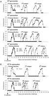
Determination of an appropriate challenge dose of P. cynomolgi sporozoites. Animals were IV challenged with varying doses of sporozoites (106, 105, 104, and 103) in PBS containing 5% BSA. The time taken for primary infection and the interval between primary infection and relapses were recorded. A dose of 106 sporozoites was used as a positive control for comparison. Parasitaemia was determined using both thick blood smears (low parasitaemia) and thin blood smears (high parasitaemia). Each parasitaemia episode was treated with CQ and for the last relapse with both CQ and PQ (A). A challenge dose of 104 sporozoites was chosen based on the results of the first experiment and was then retested in a second experiment (B)
Fig. 3
Fig. 3
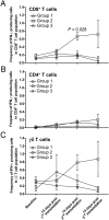
Kinetics of sporozoite-specific peripheral blood IFN-γ T cell responses. PBMC from immunized animals (groups 1 and 2) and control animals (group 3) were evaluated for the sporozoite-specific T cell responses at different time points using an ICS assay: (A) CD8+, (B) CD4+ and (C) γδ T cells. The data presented in the figure displays the mean ± SE of IFN-γ-producing cells in the CD8+, CD4+ or γδ T cell population. Individual animal data are provided in Additional file 5: Table S1
Fig. 4
Fig. 4
Kinetics of serum antibody responses. Serum samples from immunized animals (group 1 and 2) and control animals (group 3) were measured for antibody responses at different time points using IFA for both P. cynomolgi sporozoites (A) and P. cynomolgi blood-stage schizonts (B). Each data point in the graph represents the GMT with 95% CI. Individual animal data are provided in Additional file 6: Table S2
Fig. 5
Fig. 5
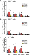
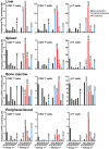
Post-challenge sporozoite-specific T cell responses in various tissue compartments. The frequencies of sporozoite-specific IFN-γ-producing T cells: (A) CD8+, (B) CD4+, and (C) γδ T cells in the liver, spleen, bone marrow, and peripheral blood were analysed by ICS assay. The data shown are means ± SE. Individual animal data are provided in Additional file 7: Table S3
Fig. 6
Fig. 6
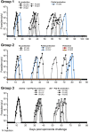
Protection induced by CPS immunization. Following challenge with IV administration of 104 sporozoites on day 112, P. cynomolgi blood-stage parasitaemia was measured in all animals until days 82–106 after the challenge using light microscopy. Each episode of parasitaemia (~ 5,000/µl) was treated with CQ for 7 days. The red lines in the graph represent parasitaemia in protected animals and the blue lines represent parasitaemia in partially protected animals
Fig. 7
Fig. 7
Potential association between T cell immune response in various tissue compartments and protection. Each bar represents the frequency of IFN-γ-producing CD8+, CD4+ and γδ T cells for each individual animal after challenge (days 194–218)

References

    1. WHO. World Malaria Report 2023. Geneva, World Health Organization; 2023. https://www.who.int/teams/global-malaria-programme/reports/world-malaria....
    1. Price RN, Tjitra E, Guerra CA, Yeung S, White NJ, Anstey NM. Vivax malaria: neglected and not benign. Am J Trop Med Hyg. 2007;77(6 Suppl):79–87. doi: 10.4269/ajtmh.2007.77.79. - DOI - PMC - PubMed
    1. Cogswell FB. The hypnozoite and relapse in primate malaria. Clin Microbiol Rev. 1992;5:26–35. doi: 10.1128/CMR.5.1.26. - DOI - PMC - PubMed
    1. Mueller I, Galinski MR, Baird JK, Carlton JM, Kochar DK, Alonso PL, et al. Key gaps in the knowledge of Plasmodium vivax, a neglected human malaria parasite. Lancet Infect Dis. 2009;9:555–566. doi: 10.1016/S1473-3099(09)70177-X. - DOI - PubMed
    1. Markus MB. Malaria: origin of the term "hypnozoite". J Hist Biol. 2011;44:781–786. doi: 10.1007/s10739-010-9239-3. - DOI - PubMed