

Nanotechnology-enabled sonodynamic therapy against malignant tumors
- PMID: 38633037
- PMCID: PMC11019498
- DOI: 10.1039/d3na00738c
Nanotechnology-enabled sonodynamic therapy against malignant tumors
Abstract
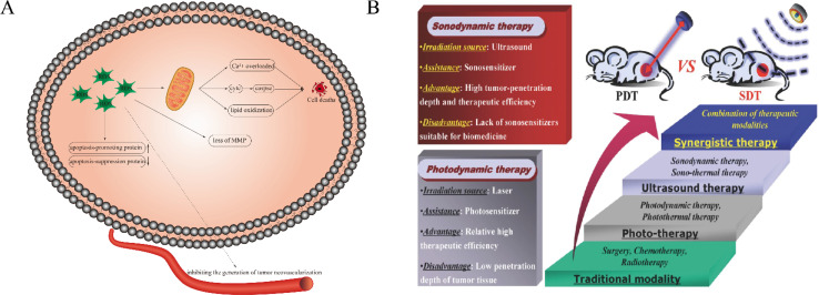
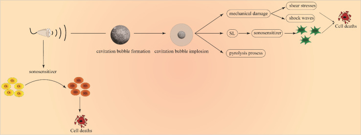
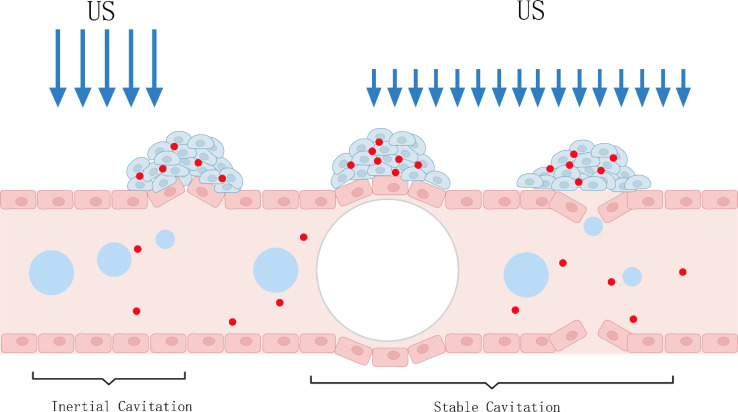
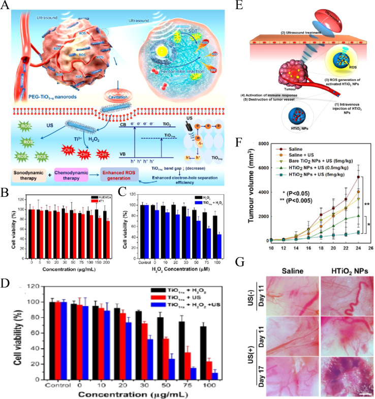
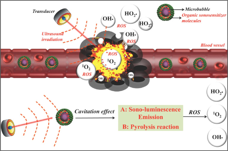
Sonodynamic therapy (SDT) is an emerging approach for malignant tumor treatment, offering high precision, deep tissue penetration, and minimal side effects. The rapid advancements in nanotechnology, particularly in cancer treatment, have enhanced the efficacy and targeting specificity of SDT. Combining sonodynamic therapy with nanotechnology offers a promising direction for future cancer treatments. In this review, we first systematically discussed the anti-tumor mechanism of SDT and then summarized the common nanotechnology-related sonosensitizers and their recent applications. Subsequently, nanotechnology-related therapies derived using the SDT mechanism were elaborated. Finally, the role of nanomaterials in SDT combined therapy was also introduced.
This journal is © The Royal Society of Chemistry.
Conflict of interest statement
The authors have declared that no competing interest exists.
Figures


Similar articles
-
A Comprehensive Review of Inorganic Sonosensitizers for Sonodynamic Therapy.Int J Mol Sci. 2023 Jul 26;24(15):12001. doi: 10.3390/ijms241512001. Int J Mol Sci. 2023. PMID: 37569377 Free PMC article. Review.
-
Inorganic Sonosensitizers for Sonodynamic Therapy in Cancer Treatment.Small. 2023 Oct;19(42):e2303195. doi: 10.1002/smll.202303195. Epub 2023 Jun 15. Small. 2023. PMID: 37323087 Review.
-
Sonodynamic therapy (SDT): a novel strategy for cancer nanotheranostics.Sci China Life Sci. 2018 Apr;61(4):415-426. doi: 10.1007/s11427-017-9262-x. Epub 2018 Apr 2. Sci China Life Sci. 2018. PMID: 29666990 Review.
-
Application of nanosonosensitizer materials in cancer sono-dynamic therapy.RSC Adv. 2022 Aug 15;12(35):22722-22747. doi: 10.1039/d2ra03786f. eCollection 2022 Aug 10. RSC Adv. 2022. PMID: 36105955 Free PMC article. Review.
-
The crosstalk between sonodynamic therapy and autophagy in cancer.Front Pharmacol. 2022 Aug 15;13:961725. doi: 10.3389/fphar.2022.961725. eCollection 2022. Front Pharmacol. 2022. PMID: 36046833 Free PMC article. Review.
Cited by
-
Exploration of Alkyne-Based Multilayered 3D Polymers and Oligomers: Subtle Aggregation-Induced Emission, Chromium(VI) Ion Detection, and Chiral Properties Characterization.Molecules. 2024 Nov 28;29(23):5641. doi: 10.3390/molecules29235641. Molecules. 2024. PMID: 39683800 Free PMC article.
-
State-of-the-art photodynamic therapy for malignant gliomas: innovations in photosensitizers and combined therapeutic approaches.Explor Target Antitumor Ther. 2025 Mar 28;6:1002303. doi: 10.37349/etat.2025.1002303. eCollection 2025. Explor Target Antitumor Ther. 2025. PMID: 40177536 Free PMC article. Review.
-
The role of manganese-based MRI contrast agents for cancer theranostics: Where do we stand in 2025?Theranostics. 2025 Mar 15;15(9):4147-4174. doi: 10.7150/thno.108705. eCollection 2025. Theranostics. 2025. PMID: 40213669 Free PMC article. Review.
-
Characteristics of Ultrasound-Driven Barium Titanate Nanoparticles and the Mechanism of Action on Solid Tumors.Int J Nanomedicine. 2024 Nov 28;19:12769-12791. doi: 10.2147/IJN.S491816. eCollection 2024. Int J Nanomedicine. 2024. PMID: 39624116 Free PMC article. Review.
-
Recent Advances and Future Directions in Sonodynamic Therapy for Cancer Treatment.BME Front. 2024 Dec 27;2024:0080. doi: 10.34133/bmef.0080. eCollection 2024. BME Front. 2024. PMID: 39735354 Free PMC article.
References
-
- Huang P. Zhang Y. Liao Y. Tang Q. Lin J. Biomimetic nanoemulsion for synergistic photodynamic‐immunotherapy against hypoxic breast tumor. Angew. Chem., Int. Ed. 2021:10647–10653. - PubMed
-
- Guan X. Yin H. A. Xu X. O. Xu G. Zhang K. Tumor Metabolism Engineered Composite Nanoplatforms Potentiate Sonodynamic Therapy via Reshaping Tumor Microenvironment and Facilitating Electron–Hole Pairs’ Separation. Adv. Funct. Mater. 2020:2000326. doi: 10.1002/adfm.202000326. - DOI
Publication types
LinkOut - more resources
Full Text Sources