. 2024 Mar 11;6(8):2198-2208.
doi: 10.1039/d3na01013a. eCollection 2024 Apr 16.

Probing the pH-dependency of DC-SIGN/R multivalent lectin-glycan interactions using polyvalent glycan-gold nanoparticles

Affiliations

Probing the pH-dependency of DC-SIGN/R multivalent lectin-glycan interactions using polyvalent glycan-gold nanoparticles

Rahman Basaran et al. Nanoscale Adv. .

Abstract

The dendritic cell tetrameric lectin, DC-SIGN, and its closely related endothelial cell lectin, DC-SIGNR (collectively abbreviated as DC-SIGN/R) play a key role in the binding and transmission of deadly viruses, including Ebola, HIV, HCV, and SARS-CoV-2. Their virus binding/release processes involve a gradually acidifying environment following the natural intracellular trafficking pathways. Therefore, understanding DC-SIGN/R's pH-dependent binding properties with glycan ligands is of great importance. We have recently developed densely glycosylated gold nanoparticles (glycan-GNPs) as a powerful new tool for probing DC-SIGN/R multivalent lectin-glycan interaction (MLGI) mechanisms. They can provide not only quantitative MLGI affinities but also important structural information, such as binding site orientation and binding modes. Herein, we further employ the glycan-GNP probes to investigate the pH dependency of DC-SIGN/R MLGI properties. We find that DC-SIGN/R MLGIs exhibit distinct pH dependence over the normal physiological (7.4) to lysosomal (∼4.6) pH range. DC-SIGN binds glycan-GNPs strongly and stably from pH 7.4 to ∼5.8, but the binding is weakened significantly as pH decreases to ≤5.4 and may be fully dissociated at pH 4.6. This behaviour is fully consistent with DC-SIGN's role as an endocytic recycling receptor. In contrast, DC-SIGNR's affinity with glycan-GNPs is enhanced with the decreasing pH from 7.4 to 5.4, peaking at pH 5.4, and then reduced as pH is further lowered. Interestingly, both DC-SIGN/R binding with glycan-GNPs are found to be partially reversible in a pH-dependent manner.


Conflict of interest statement

There are no conflicts of interest to declare.

Figures

Fig. 1
Fig. 1. (A) Schematic structure of Gx-DiMan; (B) our approach to probe DC-SIGN/R binding with Gx-DiMan via GNP fluorescence quenching: upon binding, the excited energies of fluorophore labels on DC-SIGN/R are efficiently transferred to the proximal Gx-DiMan via the NSET mechanism, leading to a greatly reduced fluorescence signal. (C) Chemical structure of the LA–EG4–DiMan ligand.
Fig. 2
Fig. 2. Fluorescence quenching efficiency (QE) – pH relationships for G13-DiMan (5.0 nM) or G27-DiMan (0.50 nM) binding with DC-SIGN (A) or DC-SIGNR (B) over a pH range of 4.6 to 7.4 at a fixed lectin : Gx molar ratio of 10 : 1 for G13-DiMan or 60 : 1 for G27-DiMan.
Fig. 3
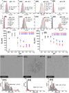
Fig. 3. (A and B) Representative Dh distribution histograms for DC-SIGN (50 nM, A1–A4) or DC-SIGNR (50 nM, B1–B4) binding with G13-DiMan (5.0 nM) at a systematically decreasing pH from 7.4 to 4.6. (C and D) Comparison of the Dh – pH relationships for DC-SIGN (C) and DC-SIGNR (D) binding with G13-DiMan (red) or G27-DiMan (blue). The Dh is displayed as mean Dh ± ½FWHM (full-width at half-maximum of the Gaussian fit). For those showing two distribution histograms, the mean Dh and mean FWHM were obtained from the linear addition of their relative abundances: i.e., mean Dh = (xc1A1%) + (xc2A2%); mean FWHM = (w1A1%) + (w2A2%). (E and F) Representative STEM images (E1–E3) and the corresponding nearest neighbour distance (NND) histograms fitted using single Gaussian distribution functions (F1–F3) of cryo-prepared G13-DiMan (5 nM) + DC-SIGN (50 nM) samples at different pHs: 7.4 (E1 and F1); 5.4 (E2 and F2), and 4.6 (E3 and F3).
Fig. 4
Fig. 4. Summary of the Dh – time dependence plots for DC-SIGN (A) or DC-SIGNR (B) binding with Gx-DiMan upon cycling the buffer pH between 7.4 and 5.0. Dhs are displayed as mean Dh ± ½FWHM obtained from the Gaussian fits. For samples displaying two distinct peaks, mean Dh = (xc1A1%) + (xc2A2%); and mean FWHM = (w1A1%) + (w2A2%).

References

    1. Huskens J., Prins L. J., Haag R. and Ravoo B. J., Multivalency: Concepts, Research and Applications, John Wiley & Sons, 2018
    1. Ribeiro-Viana R. Sánchez-Navarro M. Luczkowiak J. Koeppe J. R. Delgado R. Rojo J. Davis B. G. Nat. Commun. 2012;3:1303. doi: 10.1038/ncomms2302. - DOI - PMC - PubMed
    1. Lee R. T. Lee Y. C. Glycoconjugate J. 2000;17:543–551. doi: 10.1023/A:1011070425430. - DOI - PubMed
    1. Mammen M. Choi S. K. Whitesides G. M. Angew. Chem., Int. Ed. 1998;37:2754–2794. doi: 10.1002/(SICI)1521-3773(19981102)37:20<2754::AID-ANIE2754>3.0.CO;2-3. - DOI - PubMed
    1. Yeldell S. B. Seitz O. Chem. Soc. Rev. 2020;49:6848–6865. doi: 10.1039/D0CS00518E. - DOI - PubMed