. 2024 Mar 29;15(4):2708-2718.
doi: 10.1364/BOE.521047. eCollection 2024 Apr 1.

Removing crosstalk signals in neuron activity by time multiplexed excitations in a two-photon all-optical physiology system

Affiliations

Removing crosstalk signals in neuron activity by time multiplexed excitations in a two-photon all-optical physiology system

Chi Liu et al. Biomed Opt Express. .

Abstract

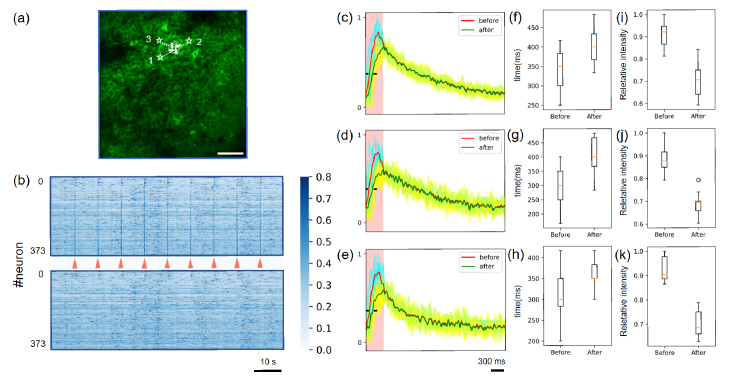
The two-photon all-optical physiology system has attracted great interest in deciphering neuronal circuits in vivo, benefiting from its advantages in recording and modulating neuronal activities at single neuron resolutions. However, the interference, or crosstalk, between the imaging and photostimulation beams introduces a significant challenge and may impede the future application of voltage indicators in two-photon all-optical physiology system. Here, we propose the time multiplexed excitation method to distinguish signals from neuronal activities and crosstalks from photostimulation. In our system, the laser pulses of the imaging beam and photostimulation beam are synchronized, and a time delay is introduced into these pulses to separate the fluorescence signal generated by these two beams. We demonstrate the efficacy of our system in eliminating crosstalk signals from photostimulation and evaluate its influence on both genetically encoded calcium indicators (GECIs) and genetically encoded voltage indicators (GEVIs) through in vivo experiments.


Conflict of interest statement

The authors declare that there are no conflicts of interest related to this article.

Figures

Fig. 1.
Fig. 1.
Signal crosstalk in two-photon all-optical physiology system. (a) Typical image of mouse hippocampus CA1 in vivo. jGCaMP8m is labeled for calcium imaging. mCherry and ChRmine are co-labeled for two-photon photostimulation. Scale bar: 80 µm. (b) Up: Emission spectra of typical red indicators. Data from [12]. Green range indicates the green filter range (500 nm ∼ 550 nm) of our two-photon all-optical physiology system. Down: Two-photon excitation spectrum for GCaMP6m. Data from [13]. Red vertical line indicates the wavelength (1040 nm) of two-photon photostimulation beam. (c) Typical images of recording channel before (t = 0 ms), during (t = 17∼51 ms) and after (t = 68, 85 ms) photostimulation. Scale bar: 80 µm.
Fig. 2.
Fig. 2.
System scheme of two-photon all-optical physiology system. A time delay of about 6.25 ns is introduced between two-photon imaging beam and two-photon stimulation beam by an optical delay line of ∼1.8 m. The excited fluorescence signals from imaging beam and stimulation beam are detected by a PMT and amplified, followed by synchronous digitization with the clock of the laser. EOM: Electric optical modulator. ETL: Electric tunable lens. BE: Beam expander. SLM: Spatial light modulator. L: Lens. SL: Scan lens. TL: Tube lens. DIC: Dichroic mirror. PMT: Photomultiplier tube.
Fig. 3.
Fig. 3.
Crosstalk signal removal in single neuron photostimulation. (a) Two-photon imaging results of recording FoV. (b) Up: ΔF/F heatmap of extracted neurons before crosstalk signal removal. Down: ΔF/F heatmap of extracted neurons after crosstalk signal removal. Orange arrowheads indicate the moment of two-photon photostimulation. (c) Correlation coefficient between other neurons and the activated neuron before and after crosstalk signal removal. (***: p < 0.001) (d) Correlation coefficient between all neurons before and after crosstalk signal removal. (***: p < 0.001) (e) Normalized ΔF/F of the activated neuron before and after crosstalk signal removal. (f) Normalized ΔF/F of a non-target neuron before and after crosstalk signal removal.
Fig. 4.
Fig. 4.
Crosstalk signal removal in serial multi-neuron photostimulation. (a) Two-photon imaging results of recording FoV. Arrows and spiral scan patterns indicate activated neurons and activation sequence. (b) Up: ΔF/F heatmap of extracted neurons before crosstalk signal removal. Down: ΔF/F heatmap of extracted neurons after crosstalk signal removal. Orange arrowheads indicate the moment of two-photon stimulation. (c-e) Zoomed-in view of neuronal activity during photostimulation of neuron #1-3. Blue and yellow trace indicate every stimuli trial before and after crosstalk signal removal. Red and green trace indicate averaged trial (N = 9) before and after crosstalk signal removal. (f-h) Time to reach the maximum activity intensity after photostimulation of neuron #1-3 before and after crosstalk signal removal. (i-k) Maximum activity intensity after photostimulation of neuron #1-3 before and after removal of signal crosstalk.
Fig. 5.
Fig. 5.
Crosstalk signal removal in holographic multi-neuron photostimulation. (a) Two-photon imaging results of recording FoV. Stars and spiral scan patterns indicate the position of activated neurons and 0th order beam. (b) Up: ΔF/F heatmap of extracted neurons before crosstalk signal removal. Down: ΔF/F heatmap of extracted neurons after crosstalk signal removal. Orange arrowheads indicate the moment of two-photon stimulation. (c-e) Zoomed-in view of neuronal activity during photostimulation of neuron #1-3. Blue and yellow trace indicate every stimuli trial before and after crosstalk signal removal. Red and green trace indicate averaged trial (N = 9) before and after crosstalk signal removal. (f-h) Time to reach the maximum activity intensity after photostimulation of neuron #1-3 before and after crosstalk signal removal. (i-k) Maximum activity intensity after photostimulation of neuron #1-3 before and after crosstalk signal removal.
Fig. 6.
Fig. 6.
Crosstalk signal removal with fast voltage indicator JEDI-2P. (a) Typical neuron expressing JEDI-2P. Scale bar: 5 µm. (b) The same neuron, as in (a), expressing jRGECO. (c) Simultaneously recording activity of the neuron in (a) and (b) in Z-score ΔF/F of JEDI-2P and jRGECO. Framerate: 207 Hz. (d) Two-photon imaging results of one FoV. Frame rate: 396.1 Hz. Spiral scan pattern and white circle indicate the stimulated neuron and a nearby neuron. (e) Two-photon imaging results of another FoV. Framerate: 396.1 Hz. Spiral excitation pattern and white circle indicate the stimulated neuron and a nearby neuron. (f) Z-score ΔF/F of the stimulated neuron [Cell 1 in (d)] before and after signal crosstalk removal. Blue and green traces indicate activity traces before and after signal crosstalk removal, respectively. Red vertical line indicates the time of photostimulation. (g) Z-score ΔF/F of the nearby neuron [Cell 2 in (d)] before and after removal of signal crosstalk removal. (h) Z-score ΔF/F of the activated neuron [Cell 1 in (e)] before and after signal crosstalk removal. (i) Z-score ΔF/F of the nearby neuron [Cell 2 in (e)] before and after signal crosstalk removal.

Similar articles

References

    1. Denk W., Strickler J. H., Webb W. W., “Two-photon laser scanning fluorescence microscopy,” Science 248(4951), 73–76 (1990).10.1126/science.2321027 - DOI - PubMed
    1. Adesnik H., Abdeladim L., “Probing neural codes with two-photon holographic optogenetics,” Nat. Neurosci. 24, 1356–1366 (2021).10.1038/s41593-021-00902-9 - DOI - PMC - PubMed
    1. Mardinly A. R., Oldenburg I. A., Pegard N. C., et al. , “Precise multimodal optical control of neural ensemble activity,” Nat. Neurosci. 21(6), 881–893 (2018).10.1038/s41593-018-0139-8 - DOI - PMC - PubMed
    1. Marshel J. H., Kim Y. S., Machado T. A., et al. , “Cortical layer–specific critical dynamics triggering perception,” Science 365(6453), eaaw5202 (2019).10.1126/science.aaw5202 - DOI - PMC - PubMed
    1. Fan L. Z., Kim D. K., Jennings J. H., et al. , “All-optical physiology resolves a synaptic basis for behavioral timescale plasticity,” Cell 136, 543–559.e519 (2023).10.1016/j.cell.2022.12.035 - DOI - PMC - PubMed

LinkOut - more resources