. 2024 Jan;14(1):512-524.
doi: 10.5455/OVJ.2024.v14.i1.46. Epub 2024 Jan 31.

Ultramicroscopic organization of the exterior olfactory organ in Anguilla vulgaris in relation to its spawning migration

Affiliations

Ultramicroscopic organization of the exterior olfactory organ in Anguilla vulgaris in relation to its spawning migration

Sheren A Al-Zahaby et al. Open Vet J. 2024 Jan.

Abstract

Background: Catadromous fishes have well-developed elongated olfactory organs with numerous lamellae and different types of receptor neurons related to their breeding migration.

Aim: The current study showed how the olfactory system adapted to the catadromous life. Our work declared the need of the migratory fishes for the sense of smell that is exhibited by a higher number of the olfactory lamellae and the receptor neuron verification in the olfactory epithelium.

Methods: Ten specimens of fully grown, but pre-matured, silver eels of Anguilla vulgaris were captured at the outlet of Edco Lake, overlooking the Mediterranean Sea, east of Alexandria. Olfactory rosettes were dissected and fixed for scanning electron microscope (SEM) and transmission electron microscope (TEM).

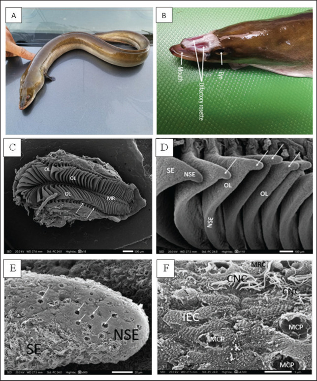
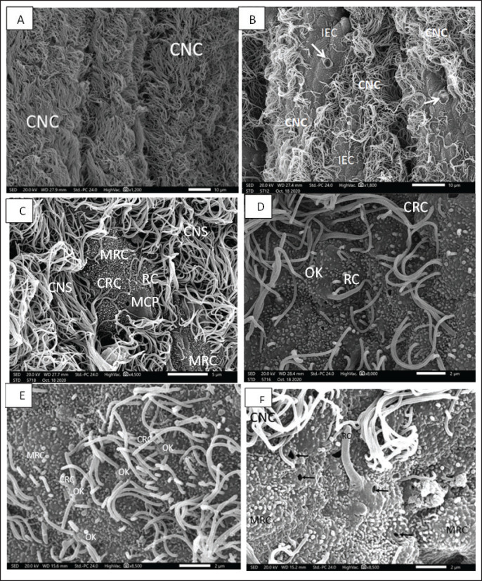
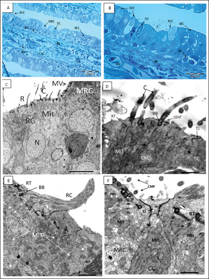
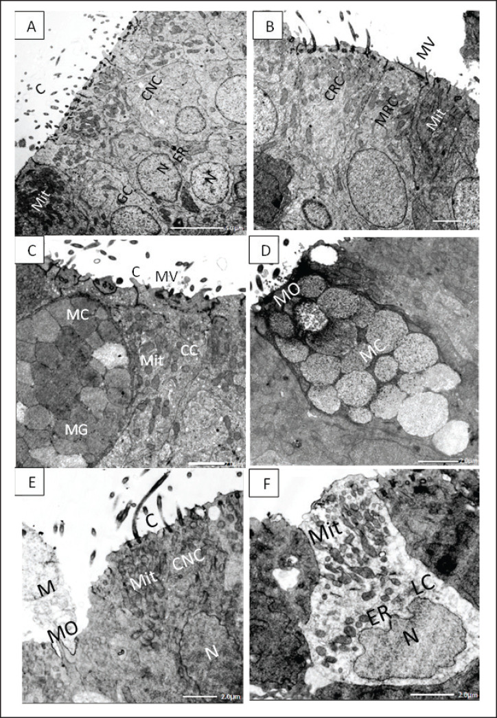
Results: Our study gave a morphological description of the olfactory system of A. vulgaris. At the ultrastructural level using SEM and TEM, one olfactory rosette was provided with 90-100 flat radial olfactory lamellae. The nasal configuration allowed water to enter and exit, transferring odorant molecules to olfactory receptor cells which comprise long cylindrical ciliated and microvillous receptors as well as rod-tipped cells. These cells are bipolar neurons with upward dendritic knobs. The olfactory epithelia also include crypt receptor cells. Interestingly, the olfactory neurons are delimited by nonsensory supporting cells, including long motile kinocilia and sustentacular supporting cells beside mucus secretory goblet cells and ionocytes or labyrinth cells that contribute to the olfaction process.

Conclusion: Olfaction is crucial in all vertebrates, including fishes as it involves reproduction, parental, feeding, defensive, schooling, and migration behaviors. Here, A. vulgaris is an excellent model for catadromous fishes. It has a well-developed olfactory organ to cope with the dramatic climate change, habitat loss, water pollution, and altered ocean currents effect during their catadromous life for reproduction.

Keywords: Anguilla vulgaris; Olfactory rosette; electron microscope; olfactory epithelium; olfactory receptors.


Conflict of interest statement

There is no conflict of interest, according to the authors.

Figures

Figure 1.
Figure 1.. A. Photograph of the whole mount (lateral view) of the studied migratory fish stage of Anguilla vulgaris. B. Photograph of the head of Anguilla vulgaris (top view) with removed skin showing the two olfactory rosette and eyes. C. Scanning Electron Micrograph (SEM) of the olfactory rosette of Anguilla vulgaris showing a whole olfactory rosette with their approximately (90–100) olfactory lamellae (OL) arranged on both sides of the median raphe (MR). Notice the connection between the wall’s capsule and lamellae (arrows). Scale bar = 500μm. D. Scanning Electron Micrograph (SEM) of the olfactory lamellae (OL) of Anguilla vulgaris showing the glandular zoon (GZ) and ciliary zoon (CZ) of nonsensory epithelium (NSE) and the sensory one (SE), linguiform process of the olfactory lamellae (arrows). Scale bar = 100μm. E. Scanning electron micrograph (SEM) of the olfactory rosette of Anguilla vulgaris showing the median central glandular zoon (GZ) and the peripheral ciliary zoon (CZ) of nonsensory epithelial area (NSE) and the sensory area (SE) in-between on the linguiform process (LP) of olfactory lamellae (OL). Scale bar = 20μm. F. SEM micrograph showing; Glandular zoon (GZ) is formed of indifferent epithelial cells (IEC) with regularly arranged microridges and wide mucous cell pores (MCP) at middle center of the linguiform process (arrow) of an olfactory lamellae (OL), however the sensory area (SE) is appeared as isolated island in-between with its ciliated receptor (CRC) and microvillous receptor (MRC). Scale bar = 5μm.
Figure 2.
Figure 2.. A. SEM micrograph showing; Strands of ciliated nonsensory cell (CNS) with their copies of tufted long cilia directed in one direction on the marginal edges of an olfactory lamella. Scale bar = 10μm. B. SEM micrograph showing; Sensory island of ciliated receptors (CRC), microvillous receptors (MRC), and rod receptors cell (RC) as well as other nonsensory island of indifferent (IEC) and mucous cell pores (MCPs) in-between copies of tufted long cilia of ciliated nonsensory cell (CNS) on an olfactory lamella. Scale bar = 10μm. C. SEM micrograph showing; two sensory islands of ciliated receptors (CRC), microvillous receptors (MRC), and rod receptors cell (RC) encountered with mucous cell pores (MCPs) in-between copies of tufted long cilia of ciliated nonsensory cell (CNS) on an olfactory lamella. Scale bar = 5μm. D. SEM micrograph of high magnification showing; sensory area of ciliated receptors (CRC), microvillous receptors (MRC), and rod-tipped receptors cell (RC) all of these dendrites emitted from a characteristic olfactory dendritic knob (OK) and encountered with mucous cell pores (MCPs) on an olfactory lamella. Scale bar = 2μm. E. SEM micrograph of high magnification showing; show; ciliated receptors cells (CRCs) with 3–5 thick somewhat short sensitive cilia and microvillous receptor cells (MRCs) with short sensitive microvilli (MV), both cilia and microvilli are emitted from a dendritic knob (OK). Scale bar = 2μm. F. SEM micrograph of high magnification showing; rod-tipped receptors cell (RC) with a very thick complex rod (R) also emitted from a dendritic knob (OK) and microvillous receptor cells (MRCs) and labyrinth cells (LCs). Scale bar = 2μm.
Figure 3.
Figure 3.. A, B Semithin section of the olfactory epithelium stained with Toluidine blue. (B) Ciliated nonsensory cells (CNCs) with cilia (C), distribution of many mucous cells (MCs), blood vessels (BVs). (C–D) The olfactory lamella consists of two layers of epithelium separated by central core (CeC) containing blood vessels (BVs) and basal lamina (BL); distribution of indifferent epithelium (IEC), olfactory receptor cell (ORC), supporting cell (SC), mucous cell (MC), basal cell (BC). C. TEM micrograph showing; Ciliated receptor (CRC) cell with short thick cilia (C), Microvillous receptor (MRC) cell with microvilli (MV), and Rod-tipped cell (RC) with compound cilium (R). All have a top mound surface, dendritic knob (OK) where their cilia, microvilli, and rod, respectively, are emitted radially. Both cells have prominent deeply positioned nucleus (N) and plentiful vesicular top-laying mitochondria (Mit). X–1,500, Scale bar: 5.0 μm. D. Higher magnification of the previous TEM micrograph showing; CRC with somewhat short thick cilia (C) emitted radially from a dendritic knob (OK), plentiful vesicular top-laying mitochondria in between two Microvillous receptor (MRC) cells. X–6,000, Scale bar: 1.0 μm. E. TEM micrograph showing; Rod-tipped receptor cell (RC) of compound cilium (R) protrudes also form a dendritic knob (OK) and has longitudinally oriented microtubules aligned in the 9 + 2 order in transverse section along its axis. This Rc is like other receptor cells has a prominent nucleus and plentiful vesicular top-laying mitochondria (Mit). X–3,000, Scale bar: 2.0 μm. F. TEM micrograph show; Crypt cell (CC) of ovoid shape bearing obscure or sunken short cilia (C) surround apical microvilli (MV). It has plentiful vesicular top-laying mitochondria (Mit) and manifested as an olfactory receptor cell. These cells are hardly detected in between the olfactory epithelia, so only are noticed in TEM figures. X–6,000, Scale bar: 1.0 μm.
Figure 4.
Figure 4.. A. TEM micrograph showing; ciliated nonsensory cells (CNS) of flat top surface and long cilia (C), deep globular nuclei with dense nucleoplasm (N), and terminal plentiful vesicular or elongated mitochondria (Mit) associated with obvious rough endoplasmic reticulum (ER) and Golgi complex (GC). X–1,500, Scale bar: 5.0 μm. B. TEM micrograph showing; ciliated receptor cell (CRC) with somewhat short thick cilia, microvillous receptor cell (MRC) with short sensory microvilli (MV) both have central prominent nucleus (N) and multiple top-laying vesicular mitochondria (Mit) and tubular endoplasmic reticulum (ER) in the labyrinth cell. X–2,000, Scale bar: 2.0 μm. C.TEM micrograph showing; mucous cell (MC) with large mucous granules (MG) in the vicinity of crypt cell (CC) bears obscure short cilia (C) surrounding apical microvilli (MV) and has plentiful vesicular top-laying mitochondria (Mit). X–2,500, Scale bar: 2.0 μm. D. TEM micrograph showing; mucous cell (MC) with large mucous granules directly opens in the olfactory surface with mucous orifice (MO). X–2,500, Scale bar: 2.0 μm. E. TEM micrograph showing; ciliated nonsensory cell (CNC) with of flat top surface and long cilia (C), central prominent nucleus (N), and plentiful vesicular mitochondria (Mit). Mucous cell orifice (MO) with discharged mucus (M). X–2,500, Scale bar: 2.0 μm. F. TEM micrograph showing; ovoid or columnar labyrinth (LC) of basely located lopsided nucleus and plentiful top-laying mitochondrial vesicles (Mit) interconnected with tubular endoplasmic reticulum (ER). X–3,000, Scale bar: 2.0 μm.

Similar articles

Cited by

References

    1. Ahuja G, Nia S.B, Zapilko V, Shiriagin V, Kowatschew D, Oka Y, Korsching S.I. Kappe neurons, a novel population of olfactory sensory neurons. Sci. Rep. 2014;4:4037. - PMC - PubMed
    1. Aicardi S, Bozzo M, Amaroli A, Gallus L, Risso B, Carlig E, Di Blasi D, Vacchi M, Ghigliotti L, Ferrando S. The arrangement of the peripheral olfactory system of Pleuragramma antarcticum: a well-exploited small sensor, an aided water flow, and a prominent effort in primary signal elaboration. Animals. 2022;12:663. - PMC - PubMed
    1. Al-Zahaby S.A, Farag M.R, Alagawany M, Taha H.S, Varoni M.V, Crescenzo G, Mawed S.A. Zinc oxide nanoparticles (ZnO-NPs) induce cytotoxicity in the zebrafish olfactory organs via activating oxidative stress and apoptosis at the ultrastructure and genetic levels. Animals. 2023;13:2867. - PMC - PubMed
    1. Atta K. Morphological, anatomical and histological studies on the olfactory organs and eyes of teleost fish: Anguilla anguilla in relation to its feeding habits. J. Basic. Appl. Zool. 2013;66:101–108.
    1. Axel R. Scents and sensibility: a molecular logic of olfactory perception (Nobel lecture) Angew. Chem. Int. Ed. Engl. 2005;44:6110–6127. - PubMed

LinkOut - more resources