. 2024 Jun 1;326(6):F1032-F1038.
doi: 10.1152/ajprenal.00051.2024. Epub 2024 Apr 18.

Commensal microbiota regulate aldosterone

Affiliations

Commensal microbiota regulate aldosterone

Brittni N Moore et al. Am J Physiol Renal Physiol. .

Abstract

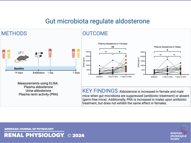
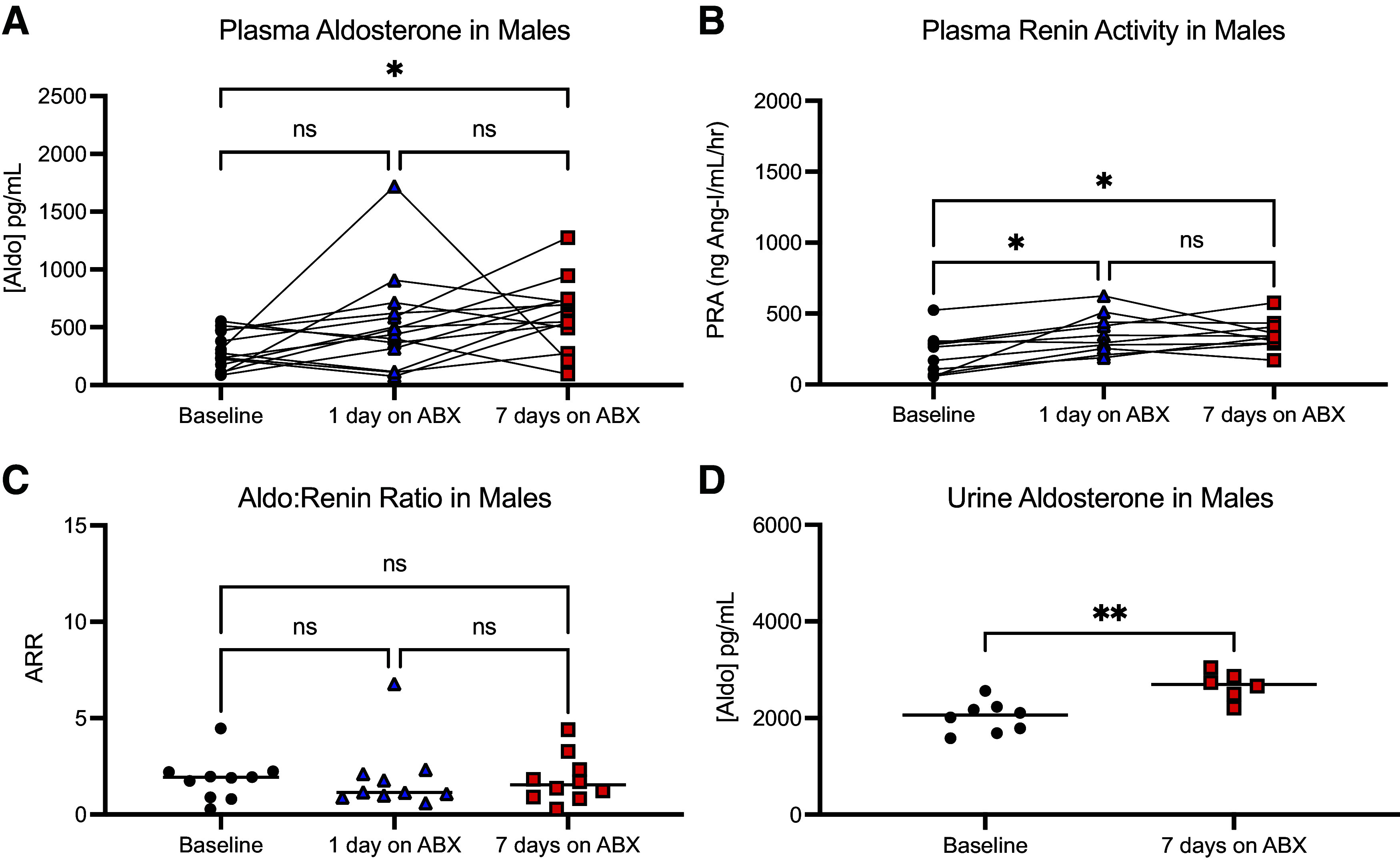
The gut microbiome regulates many important host physiological processes associated with cardiovascular health and disease; however, the impact of the gut microbiome on aldosterone is unclear. Investigating whether gut microbiota regulate aldosterone can offer novel insights into how the microbiome affects blood pressure. In this study, we aimed to determine whether gut microbiota regulate host aldosterone. We used enzyme-linked immunosorbent assays (ELISAs) to assess plasma aldosterone and plasma renin activity (PRA) in female and male mice in which gut microbiota are intact, suppressed, or absent. In addition, we examined urinary aldosterone. Our findings demonstrated that when the gut microbiota is suppressed following antibiotic treatment, there is an increase in plasma and urinary aldosterone in both female and male mice. In contrast, an increase in PRA is seen only in males. We also found that when gut microbiota are absent (germ-free mice), plasma aldosterone is significantly increased compared with conventional animals (in both females and males), but PRA is not. Understanding how gut microbiota influence aldosterone levels could provide valuable insights into the development and treatment of hypertension and/or primary aldosteronism. This knowledge may open new avenues for therapeutic interventions, such as probiotics or dietary modifications to help regulate blood pressure via microbiota-based changes to aldosterone.NEW & NOTEWORTHY We explore the role of the gut microbiome in regulating aldosterone, a hormone closely linked to blood pressure and cardiovascular disease. Despite the recognized importance of the gut microbiome in host physiology, the relationship with circulating aldosterone remains largely unexplored. We demonstrate that suppression of gut microbiota leads to increased levels of plasma and urinary aldosterone. These findings underscore the potential of the gut microbiota to influence aldosterone regulation, suggesting new possibilities for treating hypertension.

Keywords: RAAS; aldosterone; gut microbiome; renal physiology.


Conflict of interest statement

Jennifer L. Pluznick is an editor of the American Journal of Physiology-Renal Physiology and was not involved and did not have access to information regarding the peer-review process or final disposition of this article. An alternate editor oversaw the peer-review and decision-making process for this article. No conflicts of interest, financial or otherwise, are declared by the other authors.

Figures

None
Graphical abstract
Figure 1.
Figure 1.
Aldosterone and PRA in females treated with ABX. Plasma aldosterone increased after 7 days of ABX treatment in female mice (n = 16); there was also a significant increase in plasma aldosterone from day 1 to day 7 (A). No changes in plasma renin activity (PRA, n = 15, measured using the same plasma samples as shown in A, however, the n was lower due to low plasma volume for one mouse) were observed with ABX treatment (B), and, consequently, the aldosterone:renin ratio (n = 15) (C) was elevated at 7 days on ABX. Urinary aldosterone was also elevated after 7 days on ABX (D; n = 8, each sample contains urine collected from two mice). All data in this figure were from female mice; data on the same graph were from samples analyzed in the same ELISA. Identical plasma samples were used for both aldosterone and PRA experiments. Plasma aldosterone and PRA samples were analyzed using a paired one-way ANOVA with a Tukey’s post hoc test; urine aldosterone was analyzed using a nonparametric Mann–Whitney test, and ARR was analyzed using a nonparametric Kruskal–Wallis test. *P < 0.05; **P < 0.01, ***P < 0.001, ****P < 0.0001. ns, not significant.
Figure 2.
Figure 2.
Aldosterone and PRA in males treated with ABX. Plasma aldosterone increased after 7 days on ABX in male mice (n = 14) (A). Plasma renin activity significantly increased from baseline to 1 day on ABX and from baseline to 7 days on ABX (n = 10, measured using the same plasma samples as shown in A, however, the n was lower due to low plasma volume for 4 mice). There was no significant difference between 1 day on ABX and 7 days on ABX (B). As a result, there were no significant changes in the aldosterone:renin ratio (n = 10) (C). Urinary aldosterone was significantly increased from baseline to 7 days on ABX [n = 8 (baseline), 6 (7 days on ABX); each sample contained urine collected from two mice] (D). All data in this figure were from male mice; data on the same graph were from samples analyzed in the same ELISA. Identical plasma samples were used for both aldosterone and PRA experiments. Plasma aldosterone and PRA samples were analyzed using a paired one-way ANOVA with a Tukey’s post hoc test; urine aldosterone was analyzed using a nonparametric Mann–Whitney test, and ARR was analyzed using a nonparametric Kruskal–Wallis test. *P < 0.05; **P < 0.01. ns, not significant.
Figure 3.
Figure 3.
Plasma aldosterone and PRA in conventional (Conv) vs. germ-free (GF) mice. Plasma aldosterone was increased in females lacking commensal bacteria [n = 7 (Conv), 5 (GF)] (A), but plasma renin activity was unchanged [n = 7 (Conv), 5 (GF)] (B). Plasma aldosterone was significantly increased from baseline to 7 days post ABX in male mice [n = 7 (Conv), 5 (GF)] (C), but not plasma renin activity [n = 7 (Conv), 5 (GF)] (D). All data on the same graph were from samples analyzed in the same ELISA. Identical plasma samples were used for both aldosterone and PRA experiments. Data were analyzed using nonparametric Mann–Whitney tests. *P < 0.05; **P < 0.01. ns, not significant.
Figure 4.
Figure 4.
Plasma aldosterone in germ-free conventionalized (GF Conv) vs. GF mice. Plasma aldosterone was not significantly different between GF Conv (samples collected 14 days post fecal microbial transplant; n = 5) and GF (n = 5) female mice (A). Plasma aldosterone was also unchanged between males (n = 5 GF Conv and n = 5 GF) (B). All data on the same graph were from samples analyzed in the same ELISA. Data were analyzed using nonparametric Mann–Whitney tests. ns, not significant.

References

    1. Brown JM, Siddiqui M, Calhoun DA, Carey RM, Hopkins PN, Williams GH, Vaidya A. The unrecognized prevalence of primary aldosteronism: a cross-sectional study. Ann Intern Med 173: 10–20, 2020. doi:10.7326/M20-0065. - DOI - PMC - PubMed
    1. Kline GA, Prebtani APH, Leung AA, Schiffrin EL. Primary aldosteronism: a common cause of resistant hypertension. CMAJ 189: E773–E778, 2017. doi:10.1503/cmaj.161486. - DOI - PMC - PubMed
    1. Jaworska K, Koper M, Ufnal M. Gut microbiota and renin-angiotensin system: a complex interplay at local and systemic levels. Am J Physiol Gastrointest Liver Physiol 321: G355–G366, 2021. doi:10.1152/ajpgi.00099.2021. - DOI - PMC - PubMed
    1. Nakai M, Ribeiro RV, Stevens BR, Gill P, Muralitharan RR, Yiallourou S, Muir J, Carrington M, Head GA, Kaye DM, Marques FZ. Essential hypertension is associated with changes in gut microbial metabolic pathways: a multisite analysis of ambulatory blood pressure. Hypertension 78: 804–815, 2021. doi:10.1161/HYPERTENSIONAHA.121.17288. - DOI - PubMed
    1. Natarajan N, Hori D, Flavahan S, Steppan J, Flavahan NA, Berkowitz DE, Pluznick JL. Microbial short chain fatty acid metabolites lower blood pressure via endothelial G protein-coupled receptor 41. Physiol Genomics 48: 826–834, 2016. doi:10.1152/physiolgenomics.00089.2016. - DOI - PMC - PubMed

LinkOut - more resources