. 2024 Jun 1;326(6):F988-F1003.
doi: 10.1152/ajprenal.00195.2023. Epub 2024 Apr 18.

Podocyte-specific silencing of acid sphingomyelinase gene to abrogate hyperhomocysteinemia-induced NLRP3 inflammasome activation and glomerular inflammation

Affiliations

Podocyte-specific silencing of acid sphingomyelinase gene to abrogate hyperhomocysteinemia-induced NLRP3 inflammasome activation and glomerular inflammation

Dandan Huang et al. Am J Physiol Renal Physiol. .

Abstract

Acid sphingomyelinase (ASM) has been reported to increase tissue ceramide and thereby mediate hyperhomocysteinemia (hHcy)-induced glomerular nucleotide-binding oligomerization domain-like receptor containing pyrin domain 3 (NLRP3) inflammasome activation, inflammation, and sclerosis. In the present study, we tested whether somatic podocyte-specific silencing of Smpd1 gene (mouse ASM gene code) attenuates hHcy-induced NLRP3 inflammasome activation and associated extracellular vesicle (EV) release in podocytes and thereby suppresses glomerular inflammatory response and injury. In vivo, somatic podocyte-specific Smpd1 gene silencing almost blocked hHcy-induced glomerular NLRP3 inflammasome activation in Podocre (podocyte-specific expression of cre recombinase) mice compared with control littermates. By nanoparticle tracking analysis (NTA), floxed Smpd1 shRNA transfection was found to abrogate hHcy-induced elevation of urinary EV excretion in Podocre mice. In addition, Smpd1 gene silencing in podocytes prevented hHcy-induced immune cell infiltration into glomeruli, proteinuria, and glomerular sclerosis in Podocre mice. Such protective effects of podocyte-specific Smpd1 gene silencing were mimicked by global knockout of Smpd1 gene in Smpd1-/- mice. On the contrary, podocyte-specific Smpd1 gene overexpression exaggerated hHcy-induced glomerular pathological changes in Smpd1trg/Podocre (podocyte-specific Smpd1 gene overexpression) mice, which were significantly attenuated by transfection of floxed Smpd1 shRNA. In cell studies, we also confirmed that Smpd1 gene knockout or silencing prevented homocysteine (Hcy)-induced elevation of EV release in the primary cultures of podocyte isolated from Smpd1-/- mice or podocytes of Podocre mice transfected with floxed Smpd1 shRNA compared with WT/WT podocytes. Smpd1 gene overexpression amplified Hcy-induced EV secretion from podocytes of Smpd1trg/Podocre mice, which was remarkably attenuated by transfection of floxed Smpd1 shRNA. Mechanistically, Hcy-induced elevation of EV release from podocytes was blocked by ASM inhibitor (amitriptyline, AMI), but not by NLRP3 inflammasome inhibitors (MCC950 and glycyrrhizin, GLY). Super-resolution microscopy also showed that ASM inhibitor, but not NLRP3 inflammasome inhibitors, prevented the inhibition of lysosome-multivesicular body interaction by Hcy in podocytes. Moreover, we found that podocyte-derived inflammatory EVs (released from podocytes treated with Hcy) induced podocyte injury, which was exaggerated by T cell coculture. Interstitial infusion of inflammatory EVs into renal cortex induced glomerular injury and immune cell infiltration. In conclusion, our findings suggest that ASM in podocytes plays a crucial role in the control of NLRP3 inflammasome activation and inflammatory EV release during hHcy and that the development of podocyte-specific ASM inhibition or Smpd1 gene silencing may be a novel therapeutic strategy for treatment of hHcy-induced glomerular disease with minimized side effect.NEW & NOTEWORTHY In the present study, we tested whether podocyte-specific silencing of Smpd1 gene attenuates hyperhomocysteinemia (hHcy)-induced nucleotide-binding oligomerization domain-like receptor containing pyrin domain 3 (NLRP3) inflammasome activation and associated inflammatory extracellular vesicle (EV) release in podocytes and thereby suppresses glomerular inflammatory response and injury. Our findings suggest that acid sphingomyelinase (ASM) in podocytes plays a crucial role in the control of NLRP3 inflammasome activation and inflammatory EV release during hHcy. Based on our findings, it is anticipated that the development of podocyte-specific ASM inhibition or Smpd1 gene silencing may be a novel therapeutic strategy for treatment of hHcy-induced glomerular disease with minimized side effects.

Keywords: NLRP3 inflammasome; acid sphingomyelinase; extracellular vesicle; hyperhomocysteinemia; podocyte.


Conflict of interest statement

No conflicts of interest, financial or otherwise, are declared by the authors.

Figures

None
Graphical abstract
Figure 1.
Figure 1.
Characterization of in vitro and in vivo transfection of floxed Smpd1 shRNA into podocytes. A: molecular mechanism of the activation of floxed Smpd1 shRNA by cre recombinase. B: daily imaging confirmation of gene transfection by detection of cotransfected luciferase gene expression by using an in vivo imaging system (IVIS). C: representative images showing that transfection of floxed Smpd1 shRNA led to red fluorescent protein (RFP) expression in podocytes from WT/WT mice and green fluorescent protein (GFP) expression in podocytes from Podocre mice. D: representative gel documents showing the expression of acid sphingomyelinase (ASM) in podocytes from Podocre mice transfected with floxed scrambled shRNA or floxed Smpd1 shRNA. E: summarized data showing that Smpd1 gene silencing significantly decreased protein level of ASM in podocytes from Podocre mice (n = 4). F: summarized data showing that Smpd1 gene silencing significantly decreased mRNA level of ASM in podocytes from Podocre mice (n = 3–6). G: representative images showing the immunofluorescent staining of GFP expressed in Cre-positive podocytes and ASM in glomeruli of different groups of mice. H: summarized data showing that transfection of floxed Smpd1 shRNA significantly decreased ASM expression in podocytes of both Podocre and Smpd1trg/Podocre mice (n = 4). *P < 0.05 vs. the scrambled shRNA (shScra) group. #P < 0.05 vs. the Podocre group. Ctrl, control; shSmpd1, floxed Smpd1 shRNA.
Figure 2.
Figure 2.
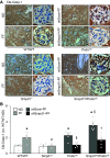
Inhibition of hyperhomocysteinemia (hHcy)-induced nucleotide-binding oligomerization domain-like receptor containing pyrin domain 3 (NLRP3) inflammasome activation in glomeruli by podocyte-specific Smpd1 gene silencing. A: representative images showing the glomerular expression of cleaved caspase-1 in different groups of mice. B: summarized data showing the glomerular expression of cleaved caspase-1 in different groups of mice (n = 6). *P < 0.05 vs. the WT/WT-normal diet (ND) group. #P < 0.05 vs. the WT/WT-folate-free (FF) group. §P < 0.05 vs. the Podocre-FF-scrambled shRNA (shScra) group. †P < 0.05 vs. the Smpd1trg/Podocre-FF-shScra group. Cle-Casp-1, cleaved caspase-1; shSmpd1, floxed Smpd1 shRNA.
Figure 3.
Figure 3.
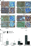
Attenuation of hyperhomocysteinemia (hHcy)-induced IL-1β production in glomeruli by podocyte-specific Smpd1 gene silencing. A: representative images showing the glomerular expression of IL-1β in different groups of mice. B: summarized data showing the glomerular expression of IL-1β in different groups of mice (n = 6). *P < 0.05 vs. the WT/WT-normal diet (ND) group. #P < 0.05 vs. the WT/WT-folate-free diet (FF) group. §P < 0.05 vs. the Podocre-FF-scrambled shRNA (shScra) group. †P < 0.05 vs. the Smpd1trg/Podocre-FF-shScra group. shSmpd1, floxed Smpd1 shRNA.
Figure 4.
Figure 4.
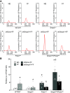
Reduction of urinary extracellular vesicle (EV) excretion by Smpd1 gene silencing in podocytes during hyperhomocysteinemia (hHcy). A: representative graphs showing urinary excretion of particles of different groups of mice detected by nanoparticle tracking analysis (NTA). B: summarized data showing urinary excretion of particles sized between 50 and 140 nm of different groups of mice (n = 7–10). *P < 0.05 vs. the WT/WT-normal diet (ND) group. #P < 0.05 vs. the WT/WT-folate-free diet (FF) group. §P < 0.05 vs. the Podocre-FF-scrambled shRNA (shScra) group. †P < 0.05 vs. the Smpd1trg/Podocre-FF-shScra group. shSmpd1, floxed Smpd1 shRNA.
Figure 5.
Figure 5.
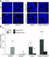
Prevention of immune cell infiltration into glomeruli by podocyte-specific Smpd1 gene silencing during hyperhomocysteinemia (hHcy). A: representative images showing the immunofluorescent staining of CD8 in glomeruli of different groups of mice. B: summarized data showing the levels of CD8 in glomeruli of different groups of mice (n = 5). *P < 0.05 vs. the WT/WT-normal diet (ND) group. #P < 0.05 vs. the WT/WT-folate-free diet (FF) group. §P < 0.05 vs. the Podocre-FF-scrambled shRNA (shScra) group. †P < 0.05 vs. the Smpd1trg/Podocre-FF-shScra group. shSmpd1, floxed Smpd1 shRNA.
Figure 6.
Figure 6.
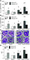
Abolishment of hyperhomocysteinemia (hHcy)-induced glomerular injury by Smpd1 gene silencing in podocytes. A: urinary protein excretion of different groups of mice (n = 7–10). B: urinary albumin excretion of different groups of mice (n = 5–8). C: representative images showing the glomerular morphological changes of different groups of mice. D: summarized data showing the glomerular damage index of different groups of mice (n = 6). *P < 0.05 vs. the WT/WT-normal diet (ND) group. #P < 0.05 vs. the WT/WT-folate-free diet (FF) group. §P < 0.05 vs. the Podocre-FF-scrambled shRNA (shScra) group. †P < 0.05 vs. the Smpd1trg/Podocre-FF-shScra group. shSmpd1, floxed Smpd1 shRNA.
Figure 7.
Figure 7.
Homocysteine (Hcy)-induced elevation of extracellular vesicle (EV) release from podocytes inhibited by Smpd1 gene silencing. A: summarized data showing EV release from different groups of podocytes (n = 6). *P < 0.05 vs. the WT/WT-vehicle (Vehl) group. #P < 0.05 vs. the WT/WT-Hcy group. §P < 0.05 vs. the Podocre-Hcy-scrambled shRNA (shScra) group. †P < 0.05 vs. the Smpd1trg/Podocre-Hcy-shScra group. Ctrl, control; shSmpd1, floxed Smpd1 shRNA. B: summarized data showing EV release from different groups of podocytes (n = 5–12). *P < 0.05 vs. the control (Ctrl) group. #P < 0.05 vs. the Vehl-Hcy group. AMI, amitriptyline; GLY, glycyrrhizin.
Figure 8.
Figure 8.
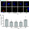
Recovery of lysosome-multivesicular body (MVB) interaction in homocysteine (Hcy)-treated podocytes by acid sphingomyelinase (ASM) inhibitor. A: representative images showing the colocalization of Rab7a and Lamp-1 in different groups of podocytes. B: summarized data showing the colocalization of Rab7a and Lamp-1 in different groups of podocytes (n = 4). *P < 0.05 vs. the control (Ctrl) group. #P < 0.05 vs. the vehicle (Vehl)-Hcy group. AMI, amitriptyline; GLY, glycyrrhizin.
Figure 9.
Figure 9.
Homocysteine (Hcy)-induced release of nucleotide-binding oligomerization domain-like receptor containing pyrin domain 3 (NLRP3) inflammasome products mediated by extracellular vesicles (EVs). A: representative gel documents and summarized data showing the levels of GM130 in podocytes and EVs (n = 4). B: representative gel documents and summarized data showing the levels of CD63 in podocytes and EVs (n = 4). *P < 0.05 vs. the podocyte group. C: summarized data of the measurement of CD63 in EVs isolated from culture medium of podocytes under control condition or treated with Hcy for 24 h by ELISA (n = 8). D: summarized data of the measurement of IL-1β in EVs isolated from culture medium of podocytes under control condition or treated with Hcy for 24 h by ELISA (n = 7). E: summarized data of the measurement of IL-18 in EVs isolated from culture medium of podocytes under control condition or treated with Hcy for 24 h by ELISA (n = 6). *P < 0.05 vs. the control (Ctrl) group.
Figure 10.
Figure 10.
Podocyte injury induced by podocyte-derived inflammatory extracellular vesicles (EVs). A: representative gel documents showing the expression of podocin in podocytes of different groups. B: summarized data showing the expression of podocin in podocytes of different groups (n = 6). C: representative gel documents showing the expression of desmin in podocytes of different groups. D: summarized data showing the expression of desmin in podocytes of different groups (n = 6). *P < 0.05 vs. the control (Ctrl) EV group. #P < 0.05 vs. the Ctrl group. Inflam EV, inflammatory EV.
Figure 11.
Figure 11.
Glomerular injury and immune cell infiltration induced by podocyte-derived inflammatory extracellular vesicles (EVs). A: representative images showing the immunofluorescent staining of podocin in glomeruli of different groups of mice. B: summarized data showing the expression of podocin in glomeruli of different groups of mice (n = 4). C: representative images showing the immunofluorescent staining of CD8 in glomeruli of different groups of mice. D: summarized data showing the levels of CD8 in glomeruli of different groups of mice (n = 4). *P < 0.05 vs. the control (Ctrl) EV group. Inflam EV, inflammatory EV.

References

    1. Li G, Kidd J, Li PL. Podocyte lysosome dysfunction in chronic glomerular diseases. Int J Mol Sci 21: 1559, 2020. doi:10.3390/ijms21051559. - DOI - PMC - PubMed
    1. Ingram AJ, Krepinsky JC, James L, Austin RC, Tang D, Salapatek AM, Thai K, Scholey JW. Activation of mesangial cell MAPK in response to homocysteine. Kidney Int 66: 733–745, 2004. doi:10.1111/j.1523-1755.2004.00795.x. - DOI - PubMed
    1. Yamasaki K, Muto J, Taylor KR, Cogen AL, Audish D, Bertin J, Grant EP, Coyle AJ, Misaghi A, Hoffman HM, Gallo RL. NLRP3/cryopyrin is necessary for interleukin-1β (IL-1β) release in response to hyaluronan, an endogenous trigger of inflammation in response to injury. J Biol Chem 284: 12762–12771, 2009. doi:10.1074/jbc.M806084200. - DOI - PMC - PubMed
    1. Martinon F, Mayor A, Tschopp J. The inflammasomes: guardians of the body. Annu Rev Immunol 27: 229–265, 2009. doi:10.1146/annurev.immunol.021908.132715. - DOI - PubMed
    1. Zhang C, Boini KM, Xia M, Abais JM, Li X, Liu Q, Li PL. Activation of Nod-like receptor protein 3 inflammasomes turns on podocyte injury and glomerular sclerosis in hyperhomocysteinemia. Hypertension 60: 154–162, 2012. doi:10.1161/HYPERTENSIONAHA.111.189688. - DOI - PMC - PubMed

MeSH terms

Substances

LinkOut - more resources