Malformations of cortical development: Fetal imaging and genetics
- PMID: 38634212
- PMCID: PMC11024634
- DOI: 10.1002/mgg3.2440
Malformations of cortical development: Fetal imaging and genetics
Abstract
Background: Malformations of cortical development (MCD) are a group of congenital disorders characterized by structural abnormalities in the brain cortex. The clinical manifestations include refractory epilepsy, mental retardation, and cognitive impairment. Genetic factors play a key role in the etiology of MCD. Currently, there is no curative treatment for MCD. Phenotypes such as epilepsy and cerebral palsy cannot be observed in the fetus. Therefore, the diagnosis of MCD is typically based on fetal brain magnetic resonance imaging (MRI), ultrasound, or genetic testing. The recent advances in neuroimaging have enabled the in-utero diagnosis of MCD using fetal ultrasound or MRI.
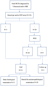
Methods: The present study retrospectively reviewed 32 cases of fetal MCD diagnosed by ultrasound or MRI. Then, the chromosome karyotype analysis, single nucleotide polymorphism array or copy number variation sequencing, and whole-exome sequencing (WES) findings were presented.
Results: Pathogenic copy number variants (CNVs) or single-nucleotide variants (SNVs) were detected in 22 fetuses (three pathogenic CNVs [9.4%, 3/32] and 19 SNVs [59.4%, 19/32]), corresponding to a total detection rate of 68.8% (22/32).
Conclusion: The results suggest that genetic testing, especially WES, should be performed for fetal MCD, in order to evaluate the outcomes and prognosis, and predict the risk of recurrence in future pregnancies.
Keywords: fetal imaging; genetics; malformations of cortical development; whole‐exome sequencing.
© 2024 The Authors. Molecular Genetics & Genomic Medicine published by Wiley Periodicals LLC.
Conflict of interest statement
All authors declare no conflicts of interest. The authors alone are responsible for the content and writing of the article.
Figures


References
-
- Accogli, A. , Severino, M. , Riva, A. , Madia, F. , Balagura, G. , Iacomino, M. , Carlini, B. , Baldassari, S. , Giacomini, T. , Croci, C. , Pisciotta, L. , Messana, T. , Boni, A. , Russo, A. , Bilo, L. , Tonziello, R. , Coppola, A. , Filla, A. , Mecarelli, O. , … Zara, F. (2020). Targeted re‐sequencing in malformations of cortical development: Genotype‐phenotype correlations. Seizure, 80, 145–152. 10.1016/j.seizure.2020.05.023 - DOI - PubMed
-
- Bedrosian, T. A. , Miller, K. E. , Grischow, O. E. , Schieffer, K. M. , LaHaye, S. , Yoon, H. , Miller, A. R. , Navarro, J. , Westfall, J. , Leraas, K. , Choi, S. , Williamson, R. , Fitch, J. , Kelly, B. J. , White, P. , Lee, K. , McGrath, S. , Cottrell, C. E. , Magrini, V. , … Koboldt, D. C. (2022). Detection of brain somatic variation in epilepsy‐associated developmental lesions. Epilepsia, 63(8), 1981–1997. 10.1111/epi.17323 - DOI - PubMed
MeSH terms
Grants and funding
- GXWCH-YMJH-2018003/the "YUMIAOJIHUA" Project of Maternal & Child Health Hospital of Guangxi Zhuang Autonomous Region
- GXWCH-ZDKF-2023-08/The open topic of Guangxi Key Laboratory of Birth Defects and Stem Cell Biobank (Maternal and Child Health Hospital of Guangxi Zhuang Autonomous Region)
- Z-A20220302/Guangxi Science and Technology Project
- grant202103000047toAY/Guangzhou Science and Technology Planning Project
LinkOut - more resources
Full Text Sources
Medical

