. 2024 Jan-Dec;16(1):2339337.
doi: 10.1080/19420862.2024.2339337. Epub 2024 Apr 18.

Reducing neonatal Fc receptor binding enhances clearance and brain-to-blood ratio of TfR-delivered bispecific amyloid-β antibody

Affiliations

Reducing neonatal Fc receptor binding enhances clearance and brain-to-blood ratio of TfR-delivered bispecific amyloid-β antibody

Eva Schlein et al. MAbs. 2024 Jan-Dec.

Abstract

Recent development of amyloid-β (Aβ)-targeted immunotherapies for Alzheimer's disease (AD) have highlighted the need for accurate diagnostic methods. Antibody-based positron emission tomography (PET) ligands are well suited for this purpose as they can be directed toward the same target as the therapeutic antibody. Bispecific, brain-penetrating antibodies can achieve sufficient brain concentrations, but their slow blood clearance remains a challenge, since it prolongs the time required to achieve a target-specific PET signal. Here, two antibodies were designed based on the Aβ antibody bapineuzumab (Bapi) - one monospecific IgG (Bapi) and one bispecific antibody with an antigen binding fragment (Fab) of the transferrin receptor (TfR) antibody 8D3 fused to one of the heavy chains (Bapi-Fab8D3) for active, TfR-mediated transport into the brain. A variant of each antibody was designed to harbor a mutation to the neonatal Fc receptor (FcRn) binding domain, to increase clearance. Blood and brain pharmacokinetics of radiolabeled antibodies were studied in wildtype (WT) and AD mice (AppNL-G-F). The FcRn mutation substantially reduced blood half-life of both Bapi and Bapi-Fab8D3. Bapi-Fab8D3 showed high brain uptake and the brain-to-blood ratio of its FcRn mutated form was significantly higher in AppNL-G-F mice than in WT mice 12 h after injection and increased further up to 168 h. Ex vivo autoradiography showed specific antibody retention in areas with abundant Aβ pathology. Taken together, these results suggest that reducing FcRn binding of a full-sized bispecific antibody increases the systemic elimination and could thereby drastically reduce the time from injection to in vivo imaging.

Keywords: Alzheimer’s disease (AD); amyloid-β (aβ); bispecific antibody; blood-brain barrier (BBB); neonatal Fc receptor (FcRn); receptor mediated transcytosis (RMT).


Conflict of interest statement

Ken G. Andersson is an employee of BioArctic AB, Sweden. The other authors have no conflicts of interest to declare.

Figures

Figure 1.
Figure 1.
Schematic visualization of the antibody design. All antibodies were based on the variable domains of Bapi in yellow, with the addition of a Fab8D3 fragment in red for bispecific variants. A black box represents mutations in the CH2, reducing the effector functions; knobs-into-holes design is visually represented by a knob and a corresponding hole; a green box represents non-mutated constant region whereas a white box indicates FcRn mutation in CH3.
Figure 2.
Figure 2.
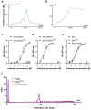
Quality control of the antibody constructs. A. Representative Biacore sensograms displaying monovalent TfR interaction of Bapi-Fab8D3 constructs. B. Representative Biacore sensograms displaying bivalent TfR interaction of 8D3 IgG. C. TfR ELISA analysis of bispecific antibody constructs D. Aβ ELISA analysis of bispecific antibody contructs E. Aβ ELISA analysis of monospecific antibody contructs F. Representative chromatogram of FcRn-column analysis of BapiFcRn- (purple), Bapi (blue) and positive control Omalizumab (orange).
Figure 3.
Figure 3.
Ex vivo analysis of antibody distribution in WT mice. A. Antibody retention in the brain, expressed as percent of injected dose per gram (%ID/g) tissue, 3 h after injection of [125I]I-Bapi, [125I]I-BapiFcRn-, [125I]I-Bapi-Fab8D3 and [125I]I-Bapi-Fab8D3FcRn- (n = 4 per antibody). B. Blood, plasma and blood cell pellet concentration (%ID/g) 3 h after injection. C. Brain-to blood ratio 3 h post injection. D. Biodistribution, expressed as organ-to-blood concentration, 3 h after injection. E. Total blood concentration (%ID/g) of the four antibodies over a time course of seven days (n = 3–4 per antibody) with two-phase decay curve fit. F. Brain concentration (%ID/g) 7 days after injection. G. Brain-to-blood ratio seven days after injection.
Figure 4.
Figure 4.
Ex vivo analysis of antibody distribution in AppNL-G-F compared to WT mice. A. Brain retention (%ID/g) of [125I]I-Bapi-Fab8D3 and [125I]I-Bapi-Fab8D3FcRn- at 12 h−168 h after injection. B. Fold difference in brain retention (AppNL-G-F/WT) at 12 h−168 h after injection of [125I]I-Bapi-Fab8D3 and [125I]I-Bapi-Fab8D3FcRn-. C. Terminal blood concentration of [125I]I-Bapi-Fab8D3 and [125I]I-Bapi-Fab8D3FcRn- in WT and AppNL-G-F mice at 12 h−168 h after injection. D. Brain-to-blood ratio of [125I]I-Bapi-Fab8D3 and [125I]I-Bapi-Fab8D3FcRn- at 12 h−168 h after injection. E. Biodistribution to peripheral organs (organ-to-blood ratio) at 24 h after injection. F. Brain retention (%ID/g) of [125I]I-Bapi and [125I]I-BapiFcRn- at 72 h after injection. G. Brain-to-blood ratio of [125I]I-Bapi and [125I]I-BapiFcRn- at 72 h after injection.
Figure 5.
Figure 5.
Post mortem analyses of antibody brain retention 24 h after injection. A. Representative images of ex vivo autoradiography illustrating the distribution of [125I]I-Bapi-Fab8D3 and [125I]I-Bapi-Fab8D3FcRn- in sagittal brain sections from AppNL-G-F and WT mice at 24 h after antibody injection. B. Immunostaining of total Aβ (3D6) in AppNL-G-F brain with squares indicating magnified areas of abundant Aβ pathology in cortex (I) and thalamus (II). C. Nuclear track emulsion autoradiography (NTE; white puncta) in combination with immunofluorescent staining of Aβ42 (green) and endothelial cell marker CD31 (pink), 24 h after injection of [125I]I-Bapi-Fab8D3FcRn- in AppNL-G-F mice, showing antibody retention along vessels and around Aβ deposits. D. Immunofluorescent staining of AppNL-G-F mouse brain with CD31 (pink) and Aβ40 (green) demonstrating abundant Aβ40 deposition along a brain vessel.

Similar articles

Cited by

References

    1. FDA . FDA grants accelerated approval for Alzheimer’s disease treatment. 2023. [accessed 2023 Jan 6 https://www.fda.gov/news-events/press-announcements/fda-grants-accelerat....
    1. Englund H, Sehlin D, Johansson A-S, Nilsson LNG, Gellerfors P, Paulie S, Lannfelt L, Pettersson FE.. Sensitive ELISA detection of amyloid-β protofibrils in biological samples. J Neurochem. 2007;103(1):334–11. doi:10.1111/j.1471-4159.2007.04759.x. - DOI - PubMed
    1. van Dyck CH, Swanson CJ, Aisen P, Bateman RJ, Chen C, Gee M, Kanekiyo M, Li D, Reyderman L, Cohen S. et al. Lecanemab in early Alzheimer’s disease. N Engl J Med. 2023;388(1):9–21. doi:10.1056/NEJMoa2212948. - DOI - PubMed
    1. Cavazzoni P. (FDA C. for D. E. and R. FDA’s decision to approve new treatment for Alzheimer’s disease. Fda’s decision to approve new treatment for Alzheimer’s disease 2021. https://www.fda.gov/drugs/news-events-human-drugs/fdas-decision-approve-....
    1. Sevigny J, Chiao P, Bussière T, Weinreb PH, Williams L, Maier M, Dunstan R, Salloway S, Chen T, Ling Y. et al. The antibody aducanumab reduces Aβ plaques in Alzheimer’s disease. Nature. 2016;537(7618):50–56. doi:10.1038/nature19323. - DOI - PubMed