

Serum GFAP levels correlate with astrocyte reactivity, post-mortem brain atrophy and neurofibrillary tangles
- PMID: 38634687
- PMCID: PMC11068326
- DOI: 10.1093/brain/awae035
Serum GFAP levels correlate with astrocyte reactivity, post-mortem brain atrophy and neurofibrillary tangles
Abstract
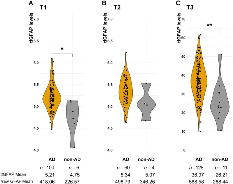
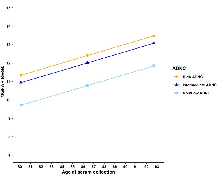
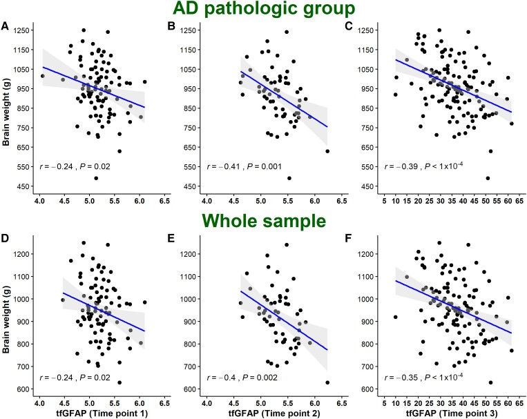
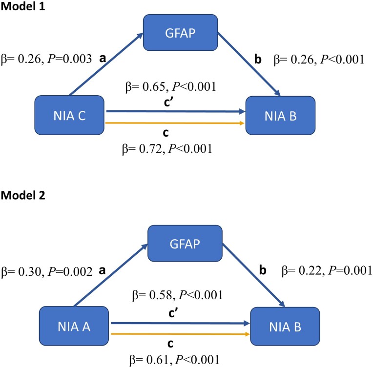
Glial fibrillary acidic protein (GFAP), a proxy of astrocyte reactivity, has been proposed as biomarker of Alzheimer's disease. However, there is limited information about the correlation between blood biomarkers and post-mortem neuropathology. In a single-centre prospective clinicopathological cohort of 139 dementia patients, for which the time-frame between GFAP level determination and neuropathological assessment was exceptionally short (on average 139 days), we analysed this biomarker, measured at three time points, in relation to proxies of disease progression such as cognitive decline and brain weight. Most importantly, we investigated the use of blood GFAP to detect the neuropathological hallmarks of Alzheimer's disease, while accounting for potential influences of the most frequent brain co-pathologies. The main findings demonstrated an association between serum GFAP level and post-mortem tau pathology (β = 12.85; P < 0.001) that was independent of amyloid deposits (β = 13.23; P = 0.02). A mediation analysis provided additional support for the role of astrocytic activation as a link between amyloid and tau pathology in Alzheimer's disease. Furthermore, a negative correlation was observed between pre-mortem serum GFAP and brain weight at post-mortem (r = -0.35; P < 0.001). This finding, together with evidence of a negative correlation with cognitive assessments (r = -0.27; P = 0.005), supports the role of GFAP as a biomarker for disease monitoring, even in the late phases of Alzheimer's disease. Moreover, the diagnostic performance of GFAP in advanced dementia patients was explored, and its discriminative power (area under the receiver operator characteristic curve at baseline = 0.91) in differentiating neuropathologically-confirmed Alzheimer's disease dementias from non-Alzheimer's disease dementias was determined, despite the challenging scenario of advanced age and frequent co-pathologies in these patients. Independently of Alzheimer's disease, serum GFAP levels were shown to be associated with two other pathologies targeting the temporal lobes-hippocampal sclerosis (β = 3.64; P = 0.03) and argyrophilic grain disease (β = -6.11; P = 0.02). Finally, serum GFAP levels were revealed to be correlated with astrocyte reactivity, using the brain GFAP-immunostained area as a proxy (ρ = 0.21; P = 0.02). Our results contribute to increasing evidence suggesting a role for blood GFAP as an Alzheimer's disease biomarker, and the findings offer mechanistic insights into the relationship between blood GFAP and Alzheimer's disease neuropathology, highlighting its ties with tau burden. Moreover, the data highlighting an independent association between serum GFAP levels and other neuropathological lesions provide information for clinicians to consider when interpreting test results. The longitudinal design and correlation with post-mortem data reinforce the robustness of our findings. However, studies correlating blood biomarkers and neuropathological assessments are still scant, and further research is needed to replicate and validate these results in diverse populations.
Keywords: Alzheimer’s disease; astrocyte; blood biomarkers; neuropathology; tau.
© The Author(s) 2024. Published by Oxford University Press on behalf of the Guarantors of Brain.
Conflict of interest statement
The authors report no competing interests.
Figures
Comment in
-
Blood GFAP reflects astrocyte reactivity to Alzheimer's pathology in post-mortem brain tissue.Brain. 2024 May 3;147(5):1598-1600. doi: 10.1093/brain/awae104. Brain. 2024. PMID: 38634672 No abstract available.
References
-
- Teunissen CE, Verberk IMW, Thijssen EH, et al. Blood-based biomarkers for Alzheimer’s disease: Towards clinical implementation. Lancet Neurol. 2022;21:66–77. - PubMed
-
- Fukuyama R, Izumoto T, Fushiki S. The cerebrospinal fluid level of glial fibrillary acidic protein is increased in cerebrospinal fluid from Alzheimer’s disease patients and correlates with severity of dementia. Eur Neurol. 2001;46:35–38. - PubMed
-
- Jesse S, Steinacker P, Cepek L, et al. Glial fibrillary acidic protein and protein S-100B: Different concentration pattern of glial proteins in cerebrospinal fluid of patients with Alzheimer’s disease and Creutzfeldt-Jakob disease. J Alzheimers Dis. 2009;17:541–551. - PubMed
-
- Ishiki A, Kamada M, Kawamura Y, et al. Glial fibrillar acidic protein in the cerebrospinal fluid of Alzheimer’s disease, dementia with Lewy bodies, and frontotemporal lobar degeneration. J Neurochem. 2016;136:258–261. - PubMed
Publication types
MeSH terms
Substances
Grants and funding
LinkOut - more resources
Full Text Sources
Medical
Miscellaneous
