. 2024 May 22;52(9):5257-5272.
doi: 10.1093/nar/gkae290.

SARS-CoV-2 nsp15 preferentially degrades AU-rich dsRNA via its dsRNA nickase activity

Affiliations

SARS-CoV-2 nsp15 preferentially degrades AU-rich dsRNA via its dsRNA nickase activity

Xionglue Wang et al. Nucleic Acids Res. .

Abstract

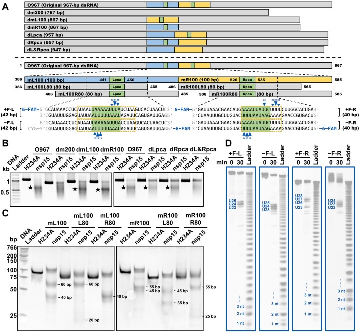
It has been proposed that coronavirus nsp15 mediates evasion of host cell double-stranded (ds) RNA sensors via its uracil-specific endoribonuclease activity. However, how nsp15 processes viral dsRNA, commonly considered as a genome replication intermediate, remains elusive. Previous research has mainly focused on short single-stranded RNA as substrates, and whether nsp15 prefers single-stranded or double-stranded RNA for cleavage is controversial. In the present work, we prepared numerous RNA substrates, including both long substrates mimicking the viral genome and short defined RNA, to clarify the substrate preference and cleavage pattern of SARS-CoV-2 nsp15. We demonstrated that SARS-CoV-2 nsp15 preferentially cleaved pyrimidine nucleotides located in less thermodynamically stable areas in dsRNA, such as AU-rich areas and mismatch-containing areas, in a nicking manner. Because coronavirus genomes generally have a high AU content, our work supported the mechanism that coronaviruses evade the antiviral response mediated by host cell dsRNA sensors by using nsp15 dsRNA nickase to directly cleave dsRNA intermediates formed during genome replication and transcription.


Figures

Graphical Abstract
Graphical Abstract
Figure 1.
Figure 1.
SARS-CoV-2 nsp15 preferentially degrades dsRNA with sequences from the SARS-CoV-2 genome. (A) Schematic showing the SARS-CoV-2 mini-genome construction. 1–265, 27 137–27 447 and 29 480–29 870 indicate the corresponding sequence ranges in the SARS-CoV-2 genome. +, − and ds refer to positive-sense ssRNA, negative-sense ssRNA, and dsRNA, respectively; TRS-CS, the core sequence of the transcription regulatory sequence; 5′ UTR (no cap), 5′ untranslated region without a 5′ cap; −M, right part of the ORF encoding the membrane protein; ORF6, the sixth ORF of the SARS-CoV-2 genome; ORF7a−, left part of the seventh ORF of the SARS-CoV-2 genome; −N, right part of the ORF encoding the nucleocapsid protein; ORF10, the last ORF of the SARS-CoV-2 genome; 3′ UTR (no polyA), 3′ untranslated region without the polyA tail. (B) Cleavage of the ssRNA and dsRNA substrates related to the SARS-CoV-2 mini-genome (as shown in A) by nsp15 in the presence of 5 mM Mn2+, 0.5 mM Mn2+ or 5 mM EDTA with various reaction times. Remaining full-length RNA substrates after nsp15 cleavage were quantified as % FL. The full gel images are shown in Supplementary Figure S6A. (C) Cleavage of the ssRNA and dsRNA substrates related to the SARS-CoV-2 mini-genome by various concentrations of nsp15 in the presence of 5 mM EDTA or 5 mM Mn2+ (0, 15, 30, 60 and 120 nM nsp15 for 5 mM EDTA; 0.125, 0.25 and 0.5 nM nsp15 for 5 mM Mn2+). (D) Cleavage of the SARS-CoV-2 mini-genome dsRNA substrates with or without TRS-CS by nsp15. In (C and D), the prominent gel bands indicating specific cleavage are marked with black pentagrams. (E) Schematic representation of RNA substrates 1–6. +, − and ds refer to positive-sense ssRNA, negative-sense ssRNA, and dsRNA, respectively. (F) Cleavage of RNA substrates 1–6 by nsp15 in the presence of 0.5 mM Mn2+. The nsp15 H234A mutant was used as a negative control. Reduction of the full-length RNA substrates by nsp15 cleavage was quantified as % ΔFL. The average and standard deviation for at least two independent reactions are graphed. The full gel images are shown in Supplementary Figure S6F. All samples were analyzed by 1% TAE AGE (native gel).
Figure 2.
Figure 2.
Identification of the cleavage sites of SARS-CoV-2 nsp15 on the dsRNA substrates derived from the SARS-CoV-2 mini-genome. (A) Schematic showing the dsRNA substrates derived from the SARS-CoV-2 mini-genome (the original 967-bp dsRNA substrate is designated as O967). Three to four sites identified with the strongest cleavage in every strand of the 6-FAM-labeled dsRNA substrates are marked with blue triangles, and the cleavage efficiency is indicated by the height of the triangle. The AU-rich areas containing these sites are marked by orange dashed boxes. Lpca, left preferred cleavage area; Rpca, right preferred cleavage area. (B) Cleavage of the O967 substrate and its variants by nsp15. Samples were analyzed by 1% TAE AGE (native gel). The prominent gel bands indicating specific cleavage are marked by black pentagrams. (C) Cleavage of the mL100 and mR100 substrates and their 80-bp variants by nsp15. Samples were analyzed by 12% TBE PAGE (native gel). The approximate sizes of the cleavage products are labeled. (D) Identification of the cleavage sites of nsp15 in the Lpca-containing and Rpca-containing short dsRNA substrates labeled with 6-FAM at the 5′ terminus. Samples were analyzed by 20% TBE-urea PAGE (denaturing gel). The three or four strongest cleavage sites in every dsRNA substrate are noted.
Figure 3.
Figure 3.
dsRNA cleavage by SARS-CoV-2 nsp15 is sensitive to AU arrangement. (A) Schematic representation of the −F-L substrate and its variants used in (B and C). The three U sites with the strongest cleavage in every strand of the −F-L substrate identified previously are shown in blue. The AU-rich areas containing nsp15 preferred cleavage sites are indicated using orange dashed boxes. The GC-rich sequences replacing the original sequences are shown in red. The black lines represent the original sequences. (B) Cleavage of the −F-L substrate and its variants as shown in (A) by nsp15. The nsp15 H234A mutant was used as a negative control. Reduction of the full-length substrate in every reaction was calculated as % ΔFL. The average and standard deviation of three independent reactions are graphed. Student's t-test was performed. ns, not significant, P > 0.05; ***P < 0.001. (C) Cleavage of the −F-L and −F-L#10 substrates by various concentrations (0, 2.5, 5, 10, 20, 40 and 80 nM) of nsp15. Reduction of the full-length substrate in every reaction was calculated as % ΔFL. In (B and C), samples were analyzed by 20% TBE PAGE (native gel). (D) Schematic representation of the mL100 substrate and its variants used in (E). The blue arrows indicate the shift direction of the dsRNA cleavage sites in the variants as compared with the mL100 substrate. (E) Shift of the dsRNA cleavage sites in the substrates shown in (D), indicated by the sizes of the cleavage products. An increased distance between the two prominent gel bands indicates a shift to the right, while a decreased distance indicates a shift to the left. (F) Schematic representation of the mL100 substrate and its variants used in (G). (G) Impact of consecutive Us on the cleavage efficiency of nsp15. (H) Schematic representation of the m100 substrate and its variant used in (I). The two UA base pairs replacing the original CG base pairs are shown in red. The AU-rich area containing consecutive Us ≥ 3 nt in both strands in the variant is marked with orange dashed boxes. (I) Enhancement of nsp15 cleavage by introduction of an AU-rich area containing consecutive Us ≥ 3 nt in both strands into dsRNA. In (G and I), the concentration of nsp15 or H234A was 10 nM. In (E, G and I), samples were analyzed by 12% TBE PAGE (native gel).
Figure 4.
Figure 4.
dsRNA thermodynamic stability and length modulate the cleavage of SARS-CoV-2 nsp15. (A) Schematic showing the +F-42.1 and −F-42.1 substrates, their variants containing a single base mismatch, and the +F-42.2 and −F-42.2 substrates. The corresponding ssRNA substrates are marked by grey dashed boxes. The preferred cleavage sites of nsp15 on the +F-42.1 and −F-42.1 substrates and their variants are shown in blue. The A base replacing the original G base in the variants of the +F-42.1 and −F-42.1 substrates are shown in red. (B) Denaturing PAGE analysis of the indicated RNA substrates. Proximity of the dsRNA gel bands to the corresponding ssRNA gel bands indicates the extent of dsRNA denaturation. (C) Thermal melting analysis of unlabeled alternatives to the +F-L, −F-L, +F-42.1v, −F-42.1v, +F-42.1, −F-42.1, +F-42.2 and −F-42.2 substrates (designated as L, 42.1v, 42.1 and 42.2). The first derivative of the melt curve produced a peak, which provided the melting temperature (Tm). RFU refers to relative fluorescence unit. (D) Cleavage of the −F-L, −F-42.1 and −F-42.2 substrates by nsp15. (E) Identification of the preferred cleavage sites of nsp15 on the +F-42.1, +F-42.1v, −F-42.1 and −F-42.1v substrates. (F) Schematic representation of the variants of the −F-L substrate with various lengths. The three U sites with the strongest cleavage in every strand of the 30-bp variants are shown in blue. The black lines represent the sequence of the −F-L30 substrates. (G) Identification of the preferred cleavage sites of nsp15 on the +F-L30 and −F-L30 substrates. The three strongest cleavage sites in every labeled strand are shown. In (E and G), samples were analyzed by 20% TBE–urea PAGE (denaturing gel). (H) Cleavage of the variants of the −F-L substrate with various lengths by nsp15. In (D and H), samples were analyzed by 20% TBE PAGE (native gel). Reduction of the full-length dsRNA caused by nsp15 cleavage was quantified as % ΔFL. The nsp15 H234A mutant was used as a negative control. The average and standard deviation for two (D) or three (H) independent reactions are graphed. Student's t-test was performed in (H). ns, not significant, P > 0.05; *P < 0.05; **P < 0.01; ****P < 0.0001.
Figure 5.
Figure 5.
SARS-CoV-2 nsp15 is a dsRNA nickase. (A) Cleavage of the 3′-6-FAM-labeled positive-sense or negative-sense strand of the dsRNA substrates shown in Supplementary Figure S2D by nsp15. Reduction of the full-length substrate in every reaction was calculated as % ΔFL. The nsp15 H234A mutant was used as a negative control. The average and standard deviation of three independent reactions are graphed. Student's t-test was performed. ***P < 0.001. (B) Cleavage of the mL100 substrate by various concentrations (0, 5, 10 and 20 nM) of nsp15. P refers to the RNA purification products of reaction samples. The gel bands above the full-length substrate gel band are marked by black arrows. (C) Schematic representation of the 40LR−15 substrate and its variants in which consecutive Us were only present in one strand within the indicated AU-rich area. (D) Cleavage of the 40LR−15 substrate and its variants shown in (C) by nsp15. The concentration of nsp15 or H234A used here was 10 nM. (E) Schematic representation of the mL100R80 substrate and its nick-containing variant. The nick is marked with a vertical blue line. (F) Cleavage of the mL100R80 substrate and its nick-containing variant by various concentrations (0, 2.5, 5 and 10 nM) of nsp15. (G) Schematic representation of the RNA-DNA hybrid substrates related to the +F-L and −F-L substrates. The DNA strands are shown in purple. (H) Cleavage of the +F-L, +F-L#, −F-L and −F-L# substrates by various concentrations (0, 5, 10 and 20 nM) of nsp15. In (A and H), samples were analyzed by 20% TBE–urea PAGE (denaturing gel). In (B, D and F), samples were analyzed by 12% TBE PAGE (native gel).
Figure 6.
Figure 6.
Coronavirus nsp15 degrades viral replication dsRNA intermediates. (A) Pie diagram grouping the coronavirus genomes based on their AU content. The genomes of 41 known coronavirus species from NCBI were included. Detailed information is listed in Supplementary Table S4. (B) Cleavage of the dsRNA substrates related to the SARS-CoV-2 mini-genome (O967) or SARS-CoV-2 S protein gene ORF (dsRNA1 and dsRNA2) into small fragments by nsp15. Samples were analyzed by 2.5% TBE AGE (native gel). The black dashed line indicates the position of the 50-bp DNA marker. (C) Schematic representation of the 39-bp 6-FAM-labeled dsRNA substrate mimicking the dsRNA replication intermediate of the 3′-polyA of the SARS-CoV-2 genome. The ssRNA substrate corresponding to the negative-sense polyU-containing strand is marked by a grey dashed box. (D) Cleavage of the polyU-containing ssRNA F-TH39− and dsRNA −F-TH39 substrates by various concentrations (0, 2.5, 5 and 10 nM) of nsp15. Samples were analyzed by 20% TBE-urea PAGE (denaturing gel). The alkaline hydrolysis products of the F-TH39− substrate were used as an RNA size ladder. (E) Model depicting how coronavirus nsp15 mediates evasion of host cell dsRNA sensors. Nsp15 preferentially cleaves consecutive Us in AU-rich areas of dsRNA in a nicking manner. The AU-rich areas are widely distributed in coronaviral genome replication dsRNA intermediates. Nsp15 efficiently cleaves dsRNA ≥40 bp, and thus is able to cleave the large dsRNA intermediates into fragments shorter than 50 bp, which may evade cytosolic dsRNA sensors, such as Mda5, PKR and OAS, even if they are released from the DMV.

References

    1. Gorbalenya A.E., Baker S.C., Baric R.S., de Groot R.J., Drosten C., Gulyaeva A.A., Haagmans B.L., Lauber C., Leontovich A.M., Neuman B.W. et al. . The species severe acute respiratory syndrome-related coronavirus: classifying 2019-nCoV and naming it SARS-CoV-2. Nat. Microbiol. 2020; 5:536–544. - PMC - PubMed
    1. Pavan M., Moro S. Lessons learnt from COVID-19: computational strategies for facing present and future pandemics. Int. J. Mol. Sci. 2023; 24:4401. - PMC - PubMed
    1. COVID-19 Genomics UK Consortium Carabelli A.M., Peacock T.P., Thorne L.G., Harvey W.T., Hughes J., Peacock S.J., Barclay W.S., de Silva T.I., Towers G.J. et al. . SARS-CoV-2 variant biology: immune escape, transmission and fitness. Nat. Rev. Micro. 2023; 21:162–177. - PMC - PubMed
    1. Huot N., Planchais C., Rosenbaum P., Contreras V., Jacquelin B., Petitdemange C., Lazzerini M., Beaumont E., Orta-Resendiz A., Rey F.A. et al. . SARS-CoV-2 viral persistence in lung alveolar macrophages is controlled by IFN-gamma and NK cells. Nat. Immunol. 2023; 24:2068–2079. - PMC - PubMed
    1. Davis H.E., McCorkell L., Vogel J.M., Topol E.J. Long COVID: major findings, mechanisms and recommendations. Nat. Rev. Micro. 2023; 21:133–146. - PMC - PubMed

Substances