. 2024 Apr 23;121(17):e2314353121.
doi: 10.1073/pnas.2314353121. Epub 2024 Apr 18.

WAV E3 ubiquitin ligases mediate degradation of IAA32/34 in the TMK1-mediated auxin signaling pathway during apical hook development

Affiliations

WAV E3 ubiquitin ligases mediate degradation of IAA32/34 in the TMK1-mediated auxin signaling pathway during apical hook development

Jun-Li Wang et al. Proc Natl Acad Sci U S A. .

Abstract

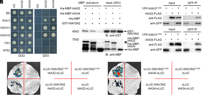
Auxin regulates plant growth and development through downstream signaling pathways, including the best-known SCFTIR1/AFB-Aux/IAA-ARF pathway and several other less characterized "noncanonical" pathways. Recently, one SCFTIR1/AFB-independent noncanonical pathway, mediated by Transmembrane Kinase 1 (TMK1), was discovered through the analyses of its functions in Arabidopsis apical hook development. Asymmetric accumulation of auxin on the concave side of the apical hook triggers DAR1-catalyzed release of the C-terminal of TMK1, which migrates into the nucleus, where it phosphorylates and stabilizes IAA32/34 to inhibit cell elongation, which is essential for full apical hook formation. However, the molecular factors mediating IAA32/34 degradation have not been identified. Here, we show that proteins in the CYTOKININ INDUCED ROOT WAVING 1 (CKRW1)/WAVY GROWTH 3 (WAV3) subfamily act as E3 ubiquitin ligases to target IAA32/34 for ubiquitination and degradation, which is inhibited by TMK1c-mediated phosphorylation. This antagonistic interaction between TMK1c and CKRW1/WAV3 subfamily E3 ubiquitin ligases regulates IAA32/34 levels to control differential cell elongation along opposite sides of the apical hook.

Keywords: Aux/IAA proteins; RING-finger E3 ubiquitin ligase; Transmembrane Kinase 1; Wavy Growth 3; noncanonical auxin signaling.


Conflict of interest statement

Competing interests statement:The authors declare no competing interest.

Figures

Fig. 1.
Fig. 1.
Loss of WAV3 E3 ubiquitin ligase activity is associated with apical hook defect. (A) Diagrams of the WAV3 protein and its truncated version. The conserved amino acids in its RING domain are shown at the Bottom. (B) Detected ubiquitin ligase activity of the truncated WAV3 in vitro. (C) C125 is essential for ubiquitin ligase activity of the WAV3 truncations. GST was used as negative control, while Coomassie Brilliant Blue (CBB) staining was the loading control. Comparable amount of GST-WAV3N2, GST-WAV3N2H147Y, and GST-WAV3N2C125S is shown in the GST-blot in SI Appendix, Fig. S4. (D) Representative apical hook phenotypes of the denoted genotypes/transgenic complementation lines 48 h after germination. Quantification of WAV3 overexpression levels is shown in SI Appendix, Fig. S5B. (E) Quantification of apical hook curvature (angle α, n ≥ 20). The box plot shows the first and third quartiles, split by the median and extended to minimum and maximum values. Different letters indicate significant differences at P < 0.05 according to one-way ANOVA with Tukey’s multiple comparisons test.
Fig. 2.
Fig. 2.
WAVs interacts with IAA32/34. (A) Yeast two-hybrid assays showing interactions between WAVs and IAA32/34. AD and BD indicate the empty vectors pGADT7 and pGBKT7, respectively. DDO/QDO denotes SD media lacking [Trp and Leu]/[Trp, Leu, His and Ade], respectively. (B) Pull-down analysis showing interactions between WAV3N2 and IAA32/34 in vitro. (C and D) Co-IP assays were used to detect the binding of WAV3C125S with IAA32 (C) or IAA34 (D). IAA32- or IAA34-FLAG was coexpressed with YFP-WAV3C125S in tobacco leaves. Isolated proteins were loaded directly on gels (input) or after IP with GFP (GFP IP). (E and F) Luciferase (LUC) complementation assays showing the interaction between WAV3N2 or its C125S mutant and IAA32 (E) or IAA34 (F) in tobacco leaves.
Fig. 3.
Fig. 3.
CKRW1/WAV3 ubiquitinates IAA32/34 for degradation. (A and B) In vitro ubiquitination of IAA32/34 by the truncated WAV3 of GST-WAV3N2, which was disrupted by the C125S mutation (C and D). The asterisks indicate ubiquitinated proteins. Comparable amount of GST-WAV3N2 and GST-WAV3N2C125S is shown in the GST-blot in SI Appendix, Fig. S4. (E and F) Comparison of overexpressed IAA32/34-GFP levels between WT (Col-0) and wav triple mutant plants. Quantification of IAA32/34-GFP bands are indicated as means ± SD (n = 3) and in SI Appendix, Fig. S9. Seedlings were treated with 100 μM cycloheximide (CHX) for the indicated time. Anti-GFP and anti-ACTIN antibodies were used for detecting the corresponding proteins. (G and H) CKRW1/WAV3 mediates IAA32/34 degradation when coexpressed in tobacco leaf cells. (Scale bars, 30 μm.) Quantification of IAA32/34-GFP fluorescence levels is shown as means ± SD (n ≥ 24) and in SI Appendix, Fig. S11.
Fig. 4.
Fig. 4.
CKRW1/WAV3-mediated degradation of IAA32/34 promotes cell elongation on the convex side of the apical hook. (A) Representative images of apical hooks in WT, wav triple mutant, and 35S:IAA32/34-GFP transgenic lines, 48 h after germination. (Scale bar, 500 μm.) Quantification of IAA32/34 overexpression levels is shown in SI Appendix, Fig. S15. (B and C) Localization of IAA32/34-GFP in etiolated seedlings of WT, wav triple and 35S:IAA32/34-GFP lines 48 h after germination. The yellow- and red-lined areas show protein levels of IAA32/34 on the concave and convex sides of the apical hook, respectively. (Scale bar, 50 μm.) Quantification of IAA32/34-GFP fluorescence density is shown as means ± SD (n ≥ 8) and in SI Appendix, Fig. S18. (D) Images of epidermal cells within the apical hook of etiolated seedlings of WT, iaa32 iaa34, wav triple, iaa32 iaa34 wav triple and 35S:IAA32/34-GFP lines, 48 h after germination. (Scale bar, 50 μm.) (E and F) Quantification of cell length. Data are mean ± SEM (n ≥ 22 in E; n ≥ 34 in F). Different uppercase or lowercase letters indicate significant differences at P < 0.05 according to one-way ANOVA with Tukey’s multiple comparisons test.
Fig. 5.
Fig. 5.
Interaction between WAVs and TMK1-mediated auxin signaling in controlling IAA32/34 stability and cell elongation in apical hook development. (A and B) Effects of WAV3 and TMK1 in IAA32/34 degradation after coexpression in tobacco leaf cells. (Scale bars, 30 μm.) Quantification of IAA32/34-GFP fluorescence levels is shown as means ± SD (n ≥ 24) and in SI Appendix, Fig. S20. (C) Images of epidermal cells within the apical hook in etiolated seedlings of WT, tmk1, wav triple and wav triple tmk1, 48 h after germination. (Scale bar, 50 μm.) (D) Quantification of cell length. Data are mean ± SEM (n ≥ 36). Different uppercase or lowercase letters indicate significant differences at P < 0.05 according to one-way ANOVA with Tukey’s multiple comparisons test. (E) Localization of IAA32/34-GFP in etiolated seedlings of tmk1 and wav triple tmk1 mutants, 48 h after germination. The yellow- and red-lined areas show the protein levels of IAA32/34 on the concave and convex sides of the apical hook, respectively. (Scale bar, 50 μm.) Quantification of IAA32/34-GFP fluorescence density is shown as means ± SD (n ≥ 7) and SI Appendix, Fig. S21.
Fig. 6.
Fig. 6.
A proposed model illustrating antagonistic interaction between the WAV ligases and the transmembrane kinase TMK1 in regulating the homeostasis of IAA32/34. IAA32/34 are noncanonical Aux/IAAs lacking the DII domain. IAA32/34 control apical hook development, possibly by interacting with ARFs. TMK1 can be cleaved by DAR1. The released kinase domain TMK1c can phosphorylate IAA32/34 to protect them from WAVs-ubiquitination-mediated proteolysis.

References

    1. Leyser O., Auxin distribution and plant pattern formation: How many angels can dance on the point of PIN? Cell 121, 819–822 (2005). - PubMed
    1. Leyser O., Auxin Signaling. Plant Physiol. 176, 465–479 (2018). - PMC - PubMed
    1. Jurado S., et al. , The Arabidopsis cell cycle F-box protein SKP2A binds to auxin. Plant Cell 22, 3891–3904 (2010). - PMC - PubMed
    1. Simonini S., et al. , A noncanonical auxin-sensing mechanism is required for organ morphogenesis in Arabidopsis. Genes. Dev. 30, 2286–2296 (2016). - PMC - PubMed
    1. Du M., Spalding E. P., Gray W. M., Rapid auxin-mediated cell expansion. Annu. Rev. Plant Biol. 71, 379–402 (2020). - PMC - PubMed

MeSH terms

LinkOut - more resources