. 2024 Jun;39(3):403-413.
doi: 10.1016/j.virs.2024.04.003. Epub 2024 Apr 16.

Pseudorabies virus manipulates mitochondrial tryptophanyl-tRNA synthetase 2 for viral replication

Affiliations

Pseudorabies virus manipulates mitochondrial tryptophanyl-tRNA synthetase 2 for viral replication

Xiu-Qing Li et al. Virol Sin. 2024 Jun.

Abstract

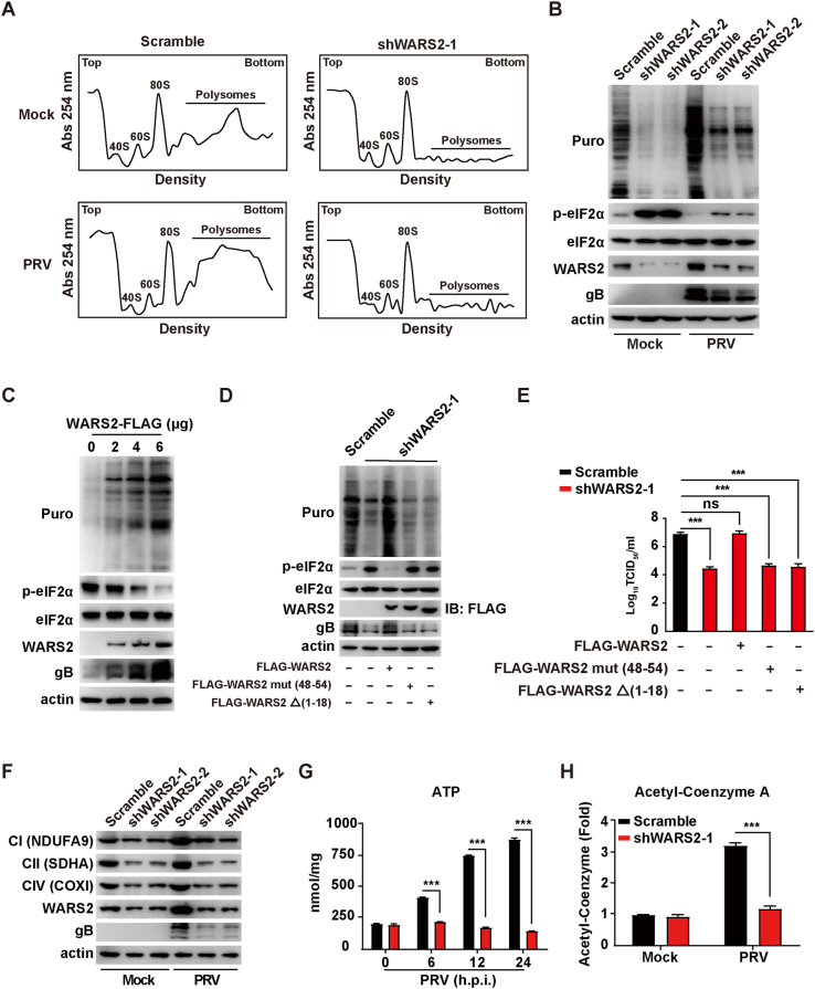
The pseudorabies virus (PRV) is identified as a double-helical DNA virus responsible for causing Aujeszky's disease, which results in considerable economic impacts globally. The enzyme tryptophanyl-tRNA synthetase 2 (WARS2), a mitochondrial protein involved in protein synthesis, is recognized for its broad expression and vital role in the translation process. The findings of our study showed an increase in both mRNA and protein levels of WARS2 following PRV infection in both cell cultures and animal models. Suppressing WARS2 expression via RNA interference in PK-15 ​cells led to a reduction in PRV infection rates, whereas enhancing WARS2 expression resulted in increased infection rates. Furthermore, the activation of WARS2 in response to PRV was found to be reliant on the cGAS/STING/TBK1/IRF3 signaling pathway and the interferon-alpha receptor-1, highlighting its regulation via the type I interferon signaling pathway. Further analysis revealed that reducing WARS2 levels hindered PRV's ability to promote protein and lipid synthesis. Our research provides novel evidence that WARS2 facilitates PRV infection through its management of protein and lipid levels, presenting new avenues for developing preventative and therapeutic measures against PRV infections.

Keywords: Innate immunity; Lipid synthesis; Protein synthesis; Pseudorabies virus; WARS2.


Conflict of interest statement

Conflict of interest The authors declare that they have no competing interests.

Figures

Fig. 1
Fig. 1
WARS2 expression is unregulated by PRV infection. A Murine models were either subjected to a mock infection or intranasal administration of PRV-QXX at a concentration of 5 ​× ​103 TCID50 within a 50 ​μL volume per specimen, maintained for a duration of 3 days. Subsequently, the expression levels of WARS2 mRNA within the renal, cerebral, pulmonary, and hepatic tissues were quantitatively analyzed via qRT-PCR (n ​= ​3 per treatment group). ∗∗∗P ​< ​0.001. B–D Following the procedural steps outlined in section (A), the expression of WARS2 and PRV glycoprotein B (gB) within the pulmonary (B), renal (C), and hepatic (D) tissues were evaluated utilizing immunoblotting analysis (n ​= ​3 per treatment group). E PK-15 ​cells were exposed to PRV-QXX with a MOI of 1 for 0–24 ​h. The expression level of WARS2 mRNA was subsequently quantified through qRT-PCR analysis. ∗∗∗P ​< ​0.001. F PK-15 ​cells were exposed to PRV-QXX with a MOI of 1 for 0–24 ​h. The protein expressions of WARS2 and gB were evaluated utilizing immunoblotting analysis. G–H PK-15 ​cells exposed to PRV-QXX with a MOI of 1 for 0–24 ​h. The expression levels of WARS1 (G) and EARS2 (H) mRNA were quantitatively analyzed via qRT-PCR.
Fig. 2
Fig. 2
WARS2 expression is dependent on type I IFN and cGAS-mediated innate immune pathway. A PK-15 ​cells were exposed to IFN-β (0–0.4 ​μg/mL) for 24 ​h. qRT-PCR analysis was employed to determine the expression levels of ISG15 and WARS2 mRNA. ∗P ​< ​0.05, ∗∗P ​< ​0.01, ∗∗∗P ​< ​0.001. B–D PK-15 ​cells underwent transfection with varying concentrations of HT-DNA (0–4 ​μg/mL) (B), poly (I:C) (0–4 ​μg/mL) (C), and poly (dA:dT) (0–4 ​μg/mL) (D) for 2 ​h. qRT-PCR analysis was conducted to quantify the mRNA expression levels of IFN-β, ISG15, and WARS2. ∗P ​< ​0.05, ∗∗P ​< ​0.01, ∗∗∗P ​< ​0.001. E The presence of cGAS, STING, TBK1, IRF3, and IFNAR1 proteins was analyzed through immunoblotting analysis in cGAS−/−, STING−/−, TBK1−/−, IRF3−/− and IFNAR1−/− PK-15 ​cells. FcGAS−/−, STING−/−, TBK1−/−, IRF3−/− and IFNAR1−/− PK-15 ​cells were subjected to infection with PRV-QXX at a MOI of 1 for 0–24 ​h. The expression level of WARS2 mRNA in these cells was quantified using qRT-PCR analysis. ∗∗∗P ​< ​0.001.
Fig. 3
Fig. 3
Suppression of WARS2 attenuates PRV infection. A WARS2 mRNA expression was quantitatively measured in scramble, shWARS2-1, and shWARS2-2 PK-15 ​cells using qRT-PCR analysis. ∗∗∗P ​< ​0.001. B Protein levels of WARS2 in PK-15 ​cells treated as described in A were determined via immunoblotting analysis. C Comparative morphology of PK-15 ​cells treated as in A was documented. Scale bar, 10 ​μm. D The viability of PK-15 ​cells with different WARS2 expression levels was evaluated over a time course of 0–60 ​h using a CCK-8 assay. E Protein levels of cGAS, STING, TBK1, IRF3, IFNAR1 and WARS2 were assessed by immunoblotting analysis in scramble, shWARS2-1, and shWARS2-2 PK-15 ​cells. F PK-15 ​cells, treated as mentioned above, were infected with PRV-GFP at an MOI of 1 for 20 ​h. GFP expression, indicative of viral replication, was monitored through fluorescence microscopy. Scale bar, 100 ​μm. G GFP-positive cells from the experiment in F were quantitated by flow cytometry. ∗∗∗P ​< ​0.001. H WARS2 and PRV gB levels were assessed by immunoblotting analysis in scramble, shWARS2-1, and shWARS2-2 PK-15 ​cells infected with PRV-QXX at an MOI of 1 for 24 ​h. I qRT-PCR analysis was employed to quantify PRV gB and TK mRNA expression in scramble, shWARS2-1, and shWARS2-2 PK-15 ​cells infected with PRV-QXX at an MOI of 1 for 24 ​h. ∗∗∗P ​< ​0.001. J Viral titers in samples from H were determined via a TCID50 assay. ∗∗∗P ​< ​0.001.
Fig. 4
Fig. 4
Enhancement of PRV infection through WARS2 overexpression. A PK-15 ​cells, post-transfection with FLAG-WARS2 plasmid (0–2 ​μg) for 12 ​h, were infected with PRV-GFP at a MOI of 1 for 20 ​h. GFP expression, as a marker of viral replication, was visualized using fluorescence microscopy. Scale bar, 100 ​μm. B Quantification of GFP-positive cells from A via flow cytometry. ∗∗P ​< ​0.01, ∗∗∗P ​< ​0.001. C Following a similar procedure as in A but with varying plasmid concentrations (0–6 ​μg), PRV gB and WARS2-FLAG protein levels were evaluated using immunoblotting analysis. D qRT-PCR analysis was utilized to assess PRV gB and TK mRNA levels in PK-15 ​cells transfected with the FLAG-WARS2 plasmid (0–2 ​μg) for 12 ​h and then infected with PRV-QXX at a MOI of 1 for 24 ​h. ∗P ​< ​0.05, ∗∗P ​< ​0.01, ∗∗∗P ​< ​0.001. E Viral replication in PK-15 ​cells treated as in D was determined via a TCID50 assay. ∗∗∗P ​< ​0.001.
Fig. 5
Fig. 5
Contribution of WARS2 to protein, ATP, and acetyl-CoA synthesis. A Scramble and shWARS2-1 PK-15 ​cells were infected with PRV-QXX at a MOI of 1 for 24 ​h. The optical density was measured at a wavelength of 254 ​nm (OD254) with varioskan flash. B Scramble, shWARS2-1 and shWARS2-2 PK-15 ​cells were mock-infected or infected with PRV-QXX at a MOI of 1 for 24 ​h, and then pulsed with 5 ​μg/mL puro for 15 ​min. Puro, p-eIF2α, eIF2α, WARS2 and PRV gB were assessed by immunoblotting analysis. C PK-15 ​cells were transfected with WARS2-FLAG (0–6 ​μg) for 24 ​h, and then pulsed with 5 ​μg/mL puro for 15 ​min. Puro, p-eIF2α, eIF2α, WARS2 and PRV gB were assessed by immunoblotting analysis. D Scramble and shWARS2-1 PK-15 ​cells were transfected with the FLAG-WARS2, FLAG-WARS2 mut (48–54) and FLAG-WARS2 Δ(1–18) plasmid 6 ​μg for 12 ​h, and infected with PRV-QXX at a MOI of 1 for 24 ​h. Puro, p-eIF2α, eIF2α, WARS2-FLAG and PRV gB were assessed by immunoblotting analysis. E Cells were infected and treated as in D. Viral titers were assessed by a TCID50 assay. ∗∗∗P ​< ​0.001. ns, no significance. F Scramble, shWARS2-1 and shWARS2-2 PK-15 ​cells were mock-infected or infected with PRV-QXX at a MOI of 1 for 24 ​h. NDUFA9 (CI), SDHA (CII), COX1 (CIV), WARS2 and gB were assessed by immunoblotting analysis. G Scramble and shWARS2-1 PK-15 ​cells were infected with PRV-QXX at a MOI of 1 for 0–24 ​h. Cellular ATP was quantified using biochemical kits. ∗∗∗P ​< ​0.001. H Scramble and shWARS2-1 PK-15 ​cells were infected with PRV-QXX at a MOI of 1 for 24 ​h. Cellular acetyl-CoA was quantified using biochemical kits. ∗∗∗P ​< ​0.001.
Fig. 6
Fig. 6
WARS2's role in PRV-induced lipid synthesis. A LDs were visualized in scramble and shWARS2-1 PK-15 ​cells post-infection with PRV-QXX at a MOI of 1 for 24 ​h using Oil Red O staining. Scale bar, 10 ​μm. B LDs quantification per cell from A was performed using ImageJ analysis (n ​= ​50). ∗∗∗P ​< ​0.001. C–E Quantification of TC (C), TG (D), and FFAs (E) in scramble and shWARS2-1 PK-15 ​cells was accomplished using biochemical kits following infection with PRV-QXX at a MOI of 1 for 0–24 ​h. ∗∗∗P ​< ​0.001. F–J mRNA expression levels of SREBP1c (F), ACACA (G), FASN (H), SREBP2 (I), and HMGCR (J) in scramble, shWARS2-1, and shWARS2-2 PK-15 ​cells were measured via qRT-PCR analysis. ∗∗∗P ​< ​0.001. K The expression of lipid metabolism enzymes and viral proteins in scramble, shWARS2-1, and shWARS2-2 PK-15 ​cells mock-infected or infected with PRV-QXX at a MOI of 1 for 24 ​h were assessed by immunoblotting analysis. L Viral titers in scramble and shWARS2-1 PK-15 ​cells, infected with PRV-QXX at a MOI of 1 and treated with oleic acid (OA at 150 ​μM) for 24 ​h, were determined through a TCID50 assay. ∗P ​< ​0.05, ∗∗P ​< ​0.01, ∗∗∗P ​< ​0.001. M–O Quantification of TC (F), TG (G), and FFAs (H) in WARS2-FLAG (0–2 ​μg) transfected PK-15 ​cells was accomplished using biochemical kits. ∗P ​< ​0.05, ∗∗∗P ​< ​0.001.
Fig. 7
Fig. 7
Schematic model of PRV manipulates mitochondrial tryptophanyl-tRNA synthetase 2 for viral replication. PRV infection activates the cGAS/STING/TBK1/IRF3 innate immune pathway, mediating type I interferon production, which in turn stimulates WARS2 expression. This enhancement in WARS2 activity boosts protein and lipid synthesis, facilitating subsequent viral replication.

Similar articles

Cited by

References

    1. Ai J.W., Weng S.S., Cheng Q., Cui P., Li Y.J., Wu H.L., Zhu Y.M., Xu B., Zhang W.H. Human endophthalmitis caused by pseudorabies virus infection, China, 2017. Emerg. Infect. Dis. 2018;24:1087–1090. - PMC - PubMed
    1. Allen C., Arjona S., Santerre M., Sawaya B. Hallmarks of metabolic reprogramming and their role in viral pathogenesis. Viruses. 2022;14:602. - PMC - PubMed
    1. Antonellis A., Green E. The role of aminoacyl-tRNA synthetases in genetic diseases. Annu. Rev. Genomics Hum. Genet. 2008;9:87–107. - PubMed
    1. Bonnevie-Nielsen V., Gerdes A., Fleckner J., Petersen J., Michelsen B., Dyrberg T. Interferon stimulates the expression of 2',5'-oligoadenylate synthetase and MHC class I antigens in insulin-producing cells. J. Interferon Res. 1991;11:255–260. - PubMed
    1. Burke E., Frucht S., Thompson K., Wolfe L., Yokoyama T., Bertoni M., Huang Y., Sincan M., Adams D., Taylor R., Gahl W., Toro C., Malicdan M. Biallelic mutations in mitochondrial tryptophanyl-tRNA synthetase cause Levodopa-responsive infantile-onset Parkinsonism. Clin. Genet. 2018;93:712–718. - PMC - PubMed

Substances

LinkOut - more resources