

Functional Connectivity Changes on Resting-State fMRI after Mild Traumatic Brain Injury: A Systematic Review
- PMID: 38637022
- PMCID: PMC11288594
- DOI: 10.3174/ajnr.A8204
Functional Connectivity Changes on Resting-State fMRI after Mild Traumatic Brain Injury: A Systematic Review
Abstract
Background: Mild traumatic brain injury is theorized to cause widespread functional changes to the brain. Resting-state fMRI may be able to measure functional connectivity changes after traumatic brain injury, but resting-state fMRI studies are heterogeneous, using numerous techniques to study ROIs across various resting-state networks.
Purpose: We systematically reviewed the literature to ascertain whether adult patients who have experienced mild traumatic brain injury show consistent functional connectivity changes on resting-state -fMRI, compared with healthy patients.
Data sources: We used 5 databases (PubMed, EMBASE, Cochrane Central, Scopus, Web of Science).
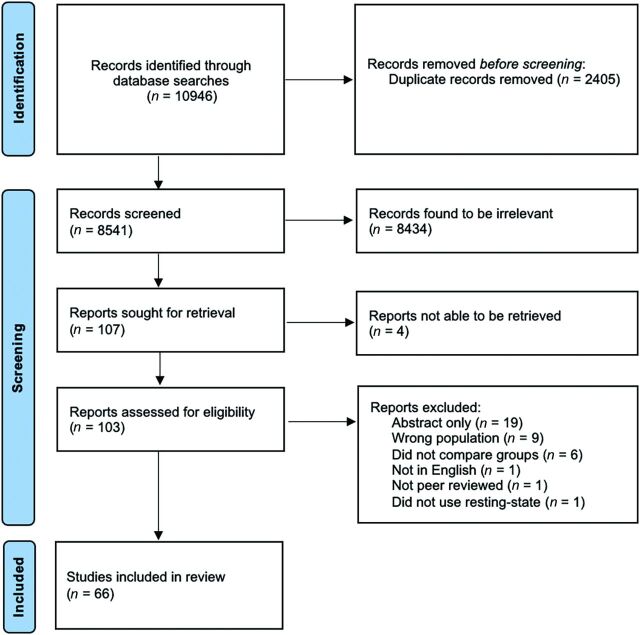
Study selection: Five databases (PubMed, EMBASE, Cochrane Central, Scopus, and Web of Science) were searched for research published since 2010. Search strategies used keywords of "functional MR imaging" and "mild traumatic brain injury" as well as related terms. All results were screened at the abstract and title levels by 4 reviewers according to predefined inclusion and exclusion criteria. For full-text inclusion, each study was evaluated independently by 2 reviewers, with discordant screening settled by consensus.
Data analysis: Data regarding article characteristics, cohort demographics, fMRI scan parameters, data analysis processing software, atlas used, data characteristics, and statistical analysis information were extracted.
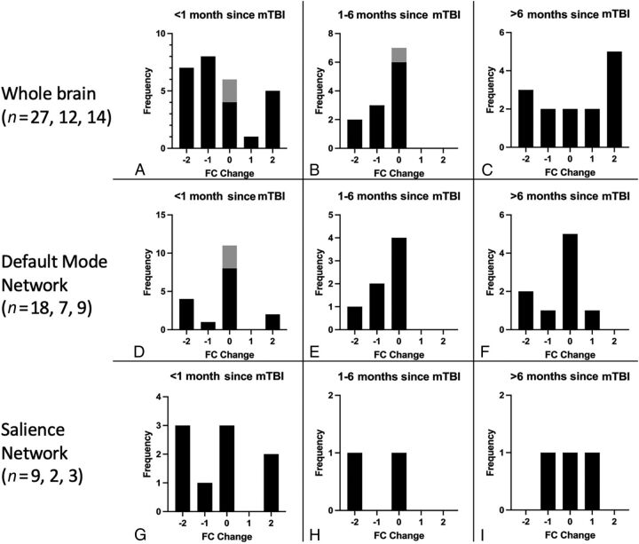
Data synthesis: Across 66 studies, 80 areas were analyzed 239 times for at least 1 time point, most commonly using independent component analysis. The most analyzed areas and networks were the whole brain, the default mode network, and the salience network. Reported functional connectivity changes varied, though there may be a slight trend toward decreased whole-brain functional connectivity within 1 month of traumatic brain injury and there may be differences based on the time since injury.
Limitations: Studies of military, sports-related traumatic brain injury, and pediatric patients were excluded. Due to the high number of relevant studies and data heterogeneity, we could not be as granular in the analysis as we would have liked.
Conclusions: Reported functional connectivity changes varied, even within the same region and network, at least partially reflecting differences in technical parameters, preprocessing software, and analysis methods as well as probable differences in individual injury. There is a need for novel rs-fMRI techniques that better capture subject-specific functional connectivity changes.
© 2024 by American Journal of Neuroradiology.
Figures
References
Publication types
MeSH terms
LinkOut - more resources
Full Text Sources
Medical