Review
. 2024 Jul;54(8):1261-1269.
doi: 10.1007/s00247-024-05911-x. Epub 2024 Apr 19.

Transseptal coronary artery-a pictorial review

Affiliations
Review

Transseptal coronary artery-a pictorial review

Vijetha V Maller et al. Pediatr Radiol. 2024 Jul.

Abstract

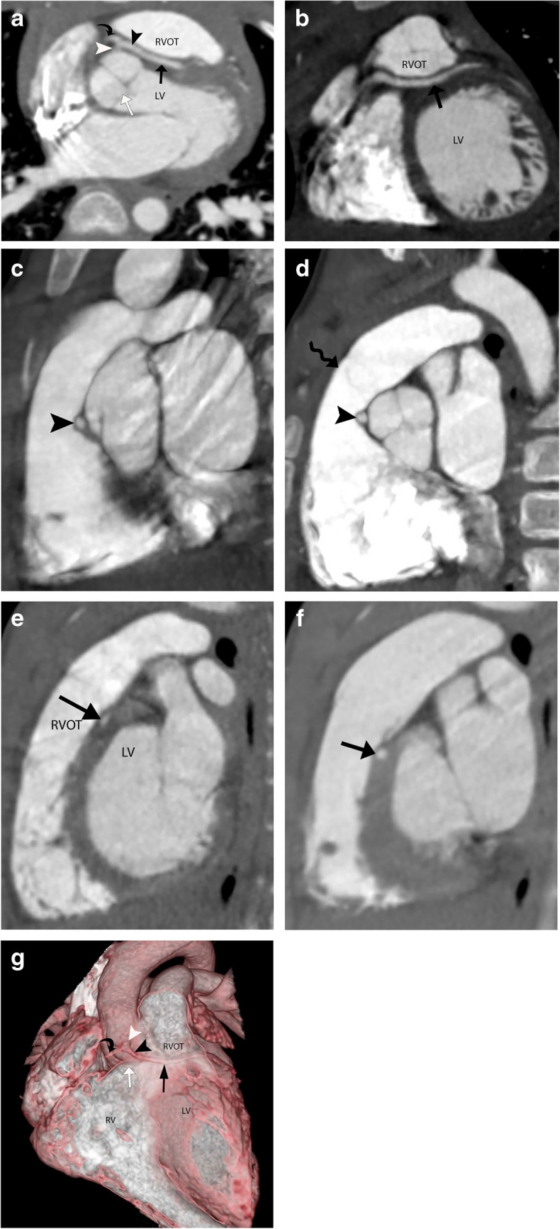
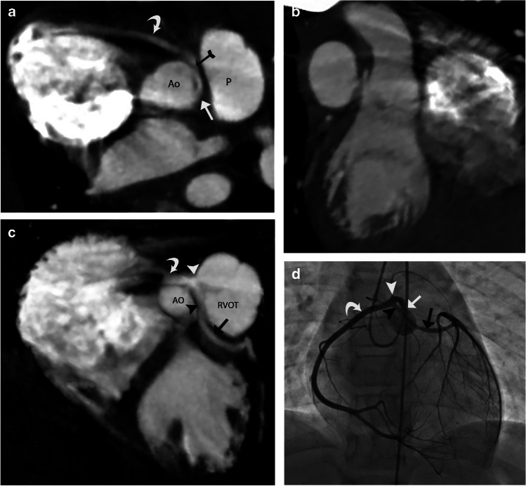
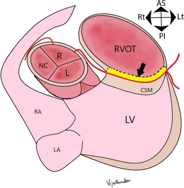
A transseptal coronary artery course, also known as a transconal course, is an anomalous course of the left main coronary artery (LMCA) or the left anterior descending artery (LAD) through the conal septal myocardium. The conal septal myocardium is the posterior wall of the right ventricular outflow tract (RVOT), acting as a dividing myocardial wall between the subaortic and subpulmonary outflow tracts. The initial segment of a transseptal coronary artery has an extraconal course between the aorta and the RVOT cranial to the true intramyocardial segment. The transseptal coronary artery then emerges out of the conal septal myocardium at the epicardial surface on the lateral aspect of the RVOT. Many consider the transseptal coronary artery to be a benign entity. However, there are few case reports of severe cardiac symptoms such as myocardial ischemia, arrhythmia, and even sudden cardiac deaths due to potential coronary artery compression in the systolic phase.​ In this article, we seek to describe the imaging findings of transseptal coronary artery course on coronary computed tomography angiography (CTA), discuss their clinical analysis, and briefly discuss the management of these lesions.

Keywords: Coronary artery; Interarterial; Intramural; Transconal; Transseptal; Unroofing; Virtual endoluminal.


Conflict of interest statement

None

Figures

Fig. 1
Fig. 1
Illustration of transseptal course of anomalous aortic origin of left main coronary artery (LMCA). Diagram in an oblique axial plane. There is an anomalous origin of LMCA from the right sinus of Valsalva (R). The LMCA then takes a sharp leftward turn as it courses between the aorta and the RVOT, which is its extraconal segment (black arrowheads). The LMCA then extends below the level of the aortic annulus (white arrow) into the posterior muscular wall of the right ventricular outflow tract (RVOT), i.e., through the conal septal myocardium (CSM), defining the transconal segment (black arrow). The LMCA, after exiting out of the transconal segment, divides into LAD (left anterior descending artery) and LCx (left circumflex artery). The RCA originates normally from the right aortic sinus of Valsalva. LV, left ventricle; LA , left atrium; RA, right atrium; R, right coronary sinus; L, left coronary sinus; NC, non-coronary sinus; AS, anterior superior; PI, posterior inferior; Rt, right; Lt, left
Fig. 2
Fig. 2
A 4-year-old asymptomatic male with a single coronary artery originating from the right sinus of Valsalva with a transseptal course of the left main coronary artery (LMCA). a Coronary CT angiography (CTA), oblique axial image. A single coronary artery (white arrowhead) originates from the right sinus of Valsalva, which then bifurcates into the right coronary artery (RCA) (black curved arrow) and LMCA (black arrowhead). The LMCA initially courses extraconally (black arrowhead) between the aorta and the right ventricular outflow tract (RVOT). The LMCA dips into the conal septum below the aortic annulus, defining the transseptal segment (black arrow). A white arrow points to the aortic annulus. b Coronary CTA, oblique coronal image. The LMCA (black arrow) courses through the conal septal myocardium with a hammock sign. LV, left ventricle. c Coronary CTA, oblique sagittal image in systolic phase (30% of RR interval). The LMCA (black arrowhead) courses extraconally between the distended aortic root and the contracted RVOT, maintaining a round caliber. d Coronary CTA, oblique sagittal image in diastolic phase (70% of RR interval). The LMCA (black arrowhead) courses extraconally between the aorta and the RVOT (below the level of pulmonary annulus shown by a wavy black arrow). The extraconal segment maintains a round caliber. e Coronary CTA oblique sagittal image in systolic phase (30% of RR interval). The LMCA (black arrow) courses through the septal myocardium between the RVOT and left ventricle (LV). The transconal LMCA (black arrow) shows an elliptical luminal caliber in the systolic phase, suggesting its myocardial compression in the systolic phase, unlike its extraconal segment. f Coronary CTA, oblique sagittal image in diastolic phase (70% of RR interval). The LMCA (black arrow) courses through the conal septal myocardium with a round luminal caliber. g 3D reformatted coronary CTA image, open view through an anterior approach. The LMCA (black arrowhead) originates from a single coronary artery (white arrowhead) arising from the right sinus of Valsalva. The extraconal segment of the LMCA (black arrowhead) extends from its origin to the level of the aortic annulus (white arrow ) between the aorta and RVOT. The intraconal segment is the hammock-like transverse segment (black arrow) below the level of the aortic annulus through the posterior wall of RVOT
Fig. 3
Fig. 3
A 17-year-old male with a cardiac murmur and anomalous aortic origin of the left anterior descending artery (LAD) from the right sinus with a transseptal course. a Coronary CT angiography (CTA), oblique axial image. LAD has an anomalous origin from the right sinus of Valsalva, after which it takes a sharp turn towards the left, coursing between the aorta and right ventricular outflow tract (RVOT). A black arrowhead points to the extraconal segment of LAD. The LAD extends into the conal septal myocardium (black arrow), defining its transconal course, and then exits out at the epicardial surface (white curved arrow). The right coronary artery (RCA) (curved black arrow) has a normal origin. LV, left ventricle. b Coronary CTA, oblique coronal image. After its anomalous origin, the LAD extends into the conal septal myocardium, defining its transconal segment (black arrow) until it extends to the epicardial surface (curved white arrow). The RCA (curved black arrow) and conus artery (white arrow) originate from the right sinus of Valsalva. LV, left ventricle. c Coronary CTA, oblique sagittal image. The anomalously originating LAD (black arrowhead) courses below the level of the pulmonary annulus (wavy black arrow) between the aorta and RVOT in the extraconal space, maintaining its round luminal caliber. There is a benign retroaortic course of the LCX (right-angled black arrow). d Coronary CTA, oblique sagittal image in systolic phase (35% of RR interval). The LAD (black arrow) courses through the conal septal myocardium, with at least 1 mm overlying septal myocardium (black chevron). Due to myocardial compression, this segment’s luminal caliber is no longer round. The retroaortic course of the LCX is also noted (right-angled black arrow). There is a tangle of vessels associated with LCX due to coronary artery fistula with the pulmonary artery in this patient (not discussed here). e Coronary CTA, endoluminal view after virtual removal of the anterior aspect of the heart and anterior wall of RVOT. The black arrowhead shows an extraconal course of the anomalously originating LAD from the opposite sinus. The block white arrow points to the entry point of the LAD into the conal septal myocardium. The LAD follows a transconal course shown by the black arrows through the conal septal myocardium (CSM). RV, right ventricle. f Coronary CTA, endoluminal view of the aortic root obtained with the camera placed in the right sinus of Valsalva pointing towards the ostium of the anomalously originating LAD from the right sinus of Valsalva. The black arrowhead points to the ostium of LAD, originating within the right sinus of Valsalva. It has a round caliber with end-on visualization of its lumen without a slitlike ostium. Five ostia are located within the right sinus of Valsalva, including the LCx (right-angled black arrow), RCA (curved black arrow), LAD (black arrowhead), and two conus arteries (white arrows), in the order of right posterior to right anterior location. A, anterior; P,  posterior; R, right; L, left. g Coronary CTA after transconal unroofing of the LAD. Oblique sagittal image in systolic phase (30% of RR interval). The anomalously originating LAD (black arrow) courses posterior to the right ventricular outflow tract (RVOT). The LAD is anterior to the conal septal myocardium (CSM), covered anteriorly by the pericardial patch (black chevron) after transconal unroofing, and hence maintains a round luminal shape in the systolic phase compared to the preoperative imaging (Fig. 3d). LV, left ventricle
Fig. 4
Fig. 4
A 9-year-old male patient who presented with cardiac arrest was noted to have a single coronary artery arising from the left sinus of Valsalva with its interarterial intramural course and additional transseptal course of the left main coronary artery (LMCA). a Coronary CT angiography (CTA), oblique axial image. A single coronary artery arises from the left sinus of Valsalva (white arrow), followed by an interarterial course between the aorta (Ao) and pulmonary artery (P) with an intramural segment. It has a hyperacute angle of origin (white arrow) and a course along the aorta. The black-tailed arrow points to the exit point of the intramural segment. The curved white arrow points to the normal course of the right coronary artery (RCA), the branch of the anomalous single coronary artery. b Coronary CTA, oblique coronal image. An elliptical luminal shape (white arrow) of the interarterial intramural single coronary artery is noted, consistent with compression. c Coronary CTA, oblique coronal image. The white arrowhead points at the bifurcation of the single coronary artery after it exits out of the intramural segment at the anterior right sinus into the RCA (curved white arrow) and LMCA (black arrowhead). The LMCA extends between the aorta (Ao) and right ventricular outflow tract (RVOT), followed by the transconal course (black arrow) as it dips into the conal septal myocardium. d Catheter angiography frontal view with selective catheter injection of the single coronary artery at the left sinus of Valsalva. The single coronary artery (white arrow), after originating from the left sinus of Valsalva, extends towards the right side, indicating its interarterial course. The white arrowhead points to the bifurcation of the single coronary artery into RCA (curved white arrow) and LMCA (black arrowhead). The LMCA then dips down like a hammock at the expected level of the RVOT, consistent with its transseptal course (black arrow). The LMCA then branches out at the lateral margin of the heart
Fig. 5
Fig. 5
Illustration of transconal unroofing of left main coronary artery (LMCA) in an oblique axial plane. The conal septal myocardium overlying the LMCA is excised (compared to Fig. 1), and the posterior wall of the right ventricular outflow tract (RVOT) is repaired with a rectangular patch of fresh autologous pericardial patch. The block black arrow points to the yellow color-coded autologous pericardium, replacing the excised portion of the septal myocardium. LV, left ventricle; LA, left atrium; RA, right atrium; R, right coronary sinus; L, left coronary sinus; NC, non-coronary sinus; AS, anterior superior; PI, posterior inferior; Rt, right; Lt, left

References

    1. Molossi S, Agrawal H, Mery CM, Krishnamurthy R, Masand P, Sexson Tejtel SK, Noel CV, Qureshi AM, Jadhav SP, McKenzie ED, Fraser CD Jr (2020) Outcomes in anomalous aortic origin of a coronary artery following a prospective standardized approach. Circ Cardiovasc Interv 13 - PubMed
    1. Najm HK, Ahmad M. Transconal unroofing of anomalous left main coronary artery from right sinus with trans-septal course. Ann Thorac Surg. 2019;108:e383–e386. doi: 10.1016/j.athoracsur.2019.04.021. - DOI - PubMed
    1. Najm HK, Karamlou T, Ahmad M, Hassan S, Salam Y, Majdalany D, Ghobrial J, Stewart RD, Unai S, Pettersson G. Early outcomes of transconal repair of transseptal anomalous left coronary artery from right sinus. Ann Thorac Surg. 2021;112:595–602. doi: 10.1016/j.athoracsur.2020.04.149. - DOI - PubMed
    1. Moore AG, Agarwal PP. Hammock sign. J Thorac Imaging. 2014;29:W89. doi: 10.1097/RTI.0000000000000095. - DOI - PubMed
    1. Brothers JA, Whitehead KK, Keller MS, Fogel MA, Paridon SM, Weinberg PM, Harris MA. Cardiac MRI and CT: differentiation of normal ostium and intraseptal course from slitlike ostium and interarterial course in anomalous left coronary artery in children. AJR Am J Roentgenol. 2015;204:W104–109. doi: 10.2214/AJR.14.12953. - DOI - PubMed

MeSH terms

LinkOut - more resources