Impact of PEG sensitization on the efficacy of PEG hydrogel-mediated tissue engineering
- PMID: 38637507
- PMCID: PMC11026400
- DOI: 10.1038/s41467-024-46327-3
Impact of PEG sensitization on the efficacy of PEG hydrogel-mediated tissue engineering
Abstract
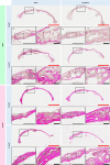
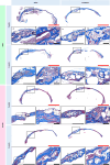
While poly(ethylene glycol) (PEG) hydrogels are generally regarded as biologically inert blank slates, concerns over PEG immunogenicity are growing, and the implications for tissue engineering are unknown. Here, we investigate these implications by immunizing mice against PEG to stimulate anti-PEG antibody production and evaluating bone defect regeneration after treatment with bone morphogenetic protein-2-loaded PEG hydrogels. Quantitative analysis reveals that PEG sensitization increases bone formation compared to naive controls, whereas histological analysis shows that PEG sensitization induces an abnormally porous bone morphology at the defect site, particularly in males. Furthermore, immune cell recruitment is higher in PEG-sensitized mice administered the PEG-based treatment than their naive counterparts. Interestingly, naive controls that were administered a PEG-based treatment also develop anti-PEG antibodies. Sex differences in bone formation and immune cell recruitment are also apparent. Overall, these findings indicate that anti-PEG immune responses can impact tissue engineering efficacy and highlight the need for further investigation.
© 2024. The Author(s).
Conflict of interest statement
The authors declare no competing interests.
Figures


Similar articles
-
Incorporation of a silicon-based polymer to PEG-DA templated hydrogel scaffolds for bioactivity and osteoinductivity.Acta Biomater. 2019 Nov;99:100-109. doi: 10.1016/j.actbio.2019.09.018. Epub 2019 Sep 16. Acta Biomater. 2019. PMID: 31536841 Free PMC article.
-
Click-based injectable bioactive PEG-hydrogels guide rapid craniomaxillofacial bone regeneration by the spatiotemporal delivery of rhBMP-2.J Mater Chem B. 2023 Apr 5;11(14):3136-3150. doi: 10.1039/d2tb02703h. J Mater Chem B. 2023. PMID: 36896831
-
Early biocompatibility of poly (ethylene glycol) hydrogel barrier materials for guided bone regeneration. An in vitro study using human gingival fibroblasts (HGF-1).Clin Oral Implants Res. 2014 Jan;25(1):16-20. doi: 10.1111/clr.12076. Epub 2012 Nov 23. Clin Oral Implants Res. 2014. PMID: 23173910
-
Fabrication of tough poly(ethylene glycol)/collagen double network hydrogels for tissue engineering.J Biomed Mater Res A. 2018 Jan;106(1):192-200. doi: 10.1002/jbm.a.36222. Epub 2017 Sep 28. J Biomed Mater Res A. 2018. PMID: 28884502
-
A review of emerging bone tissue engineering via PEG conjugated biodegradable amphiphilic copolymers.Mater Sci Eng C Mater Biol Appl. 2019 Apr;97:1021-1035. doi: 10.1016/j.msec.2019.01.057. Epub 2019 Jan 15. Mater Sci Eng C Mater Biol Appl. 2019. PMID: 30678893 Review.
Cited by
-
Nonviral targeted mRNA delivery: principles, progresses, and challenges.MedComm (2020). 2025 Jan 2;6(1):e70035. doi: 10.1002/mco2.70035. eCollection 2025 Jan. MedComm (2020). 2025. PMID: 39760110 Free PMC article. Review.
-
Recent Advances in Polyurethane for Artificial Vascular Application.Polymers (Basel). 2024 Dec 18;16(24):3528. doi: 10.3390/polym16243528. Polymers (Basel). 2024. PMID: 39771380 Free PMC article. Review.
-
Highly tough and conformal silk-based adhesive patches for sutureless repair of gastrointestinal and peripheral nerve defects.Bioact Mater. 2025 Jul 4;53:1-19. doi: 10.1016/j.bioactmat.2025.06.057. eCollection 2025 Nov. Bioact Mater. 2025. PMID: 40688023 Free PMC article.
-
New Trends in Preparation and Use of Hydrogels for Water Treatment.Gels. 2025 Mar 24;11(4):238. doi: 10.3390/gels11040238. Gels. 2025. PMID: 40277674 Free PMC article. Review.
-
Advancements in Hydrogels: A Comprehensive Review of Natural and Synthetic Innovations for Biomedical Applications.Polymers (Basel). 2025 Jul 24;17(15):2026. doi: 10.3390/polym17152026. Polymers (Basel). 2025. PMID: 40808075 Free PMC article. Review.
References
MeSH terms
Substances
Grants and funding
LinkOut - more resources
Full Text Sources

