. 2024 Apr 18;14(1):8998.
doi: 10.1038/s41598-024-59451-3.

Disruption of the pro-oncogenic c-RAF-PDE8A complex represents a differentiated approach to treating KRAS-c-RAF dependent PDAC

Affiliations

Disruption of the pro-oncogenic c-RAF-PDE8A complex represents a differentiated approach to treating KRAS-c-RAF dependent PDAC

Sean F Cooke et al. Sci Rep. .

Abstract

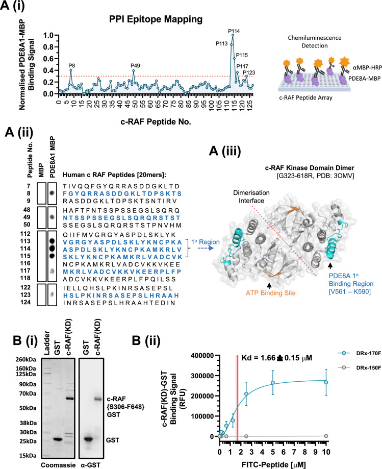
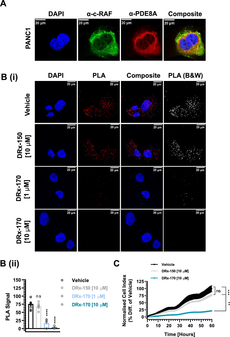
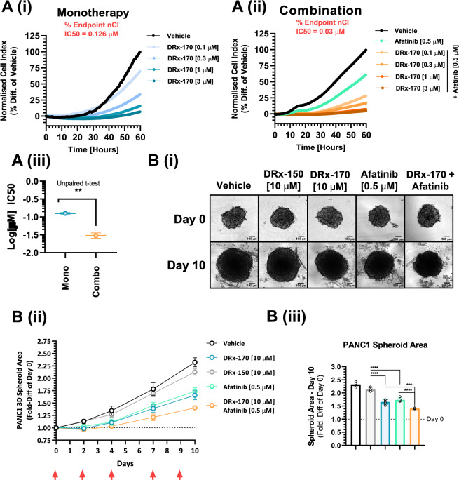
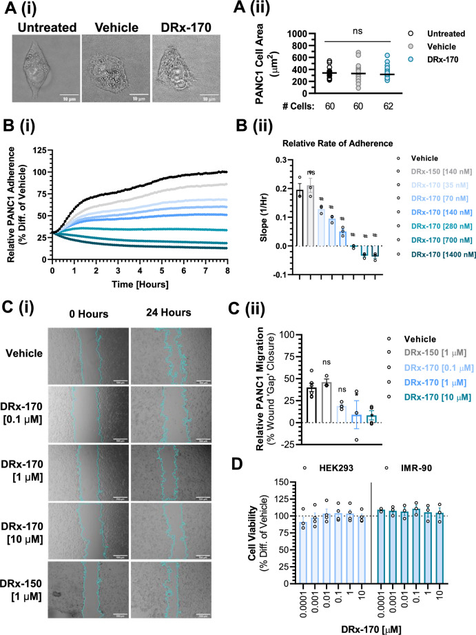
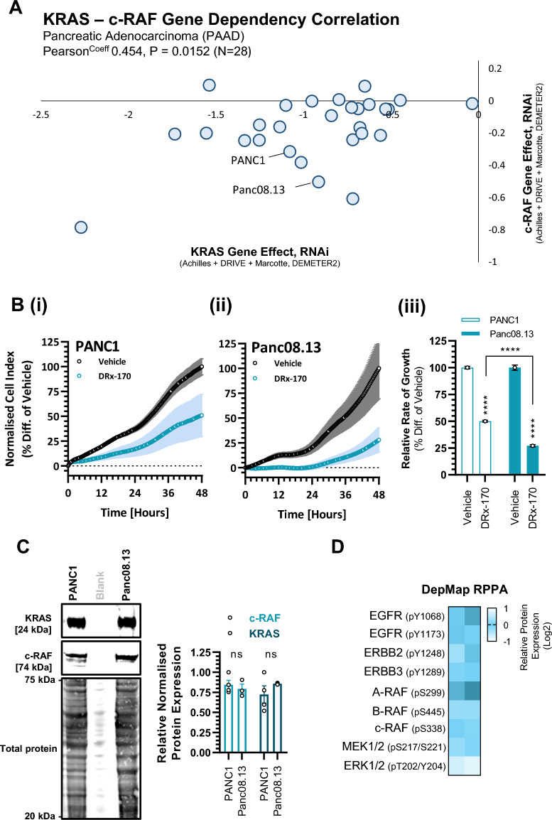
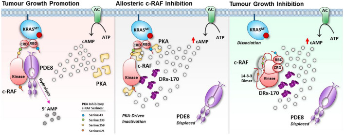
Pancreatic ductal adenocarcinoma (PDAC) is considered the third leading cause of cancer mortality in the western world, offering advanced stage patients with few viable treatment options. Consequently, there remains an urgent unmet need to develop novel therapeutic strategies that can effectively inhibit pro-oncogenic molecular targets underpinning PDACs pathogenesis and progression. One such target is c-RAF, a downstream effector of RAS that is considered essential for the oncogenic growth and survival of mutant RAS-driven cancers (including KRASMT PDAC). Herein, we demonstrate how a novel cell-penetrating peptide disruptor (DRx-170) of the c-RAF-PDE8A protein-protein interaction (PPI) represents a differentiated approach to exploiting the c-RAF-cAMP/PKA signaling axes and treating KRAS-c-RAF dependent PDAC. Through disrupting the c-RAF-PDE8A protein complex, DRx-170 promotes the inactivation of c-RAF through an allosteric mechanism, dependent upon inactivating PKA phosphorylation. DRx-170 inhibits cell proliferation, adhesion and migration of a KRASMT PDAC cell line (PANC1), independent of ERK1/2 activity. Moreover, combining DRx-170 with afatinib significantly enhances PANC1 growth inhibition in both 2D and 3D cellular models. DRx-170 sensitivity appears to correlate with c-RAF dependency. This proof-of-concept study supports the development of DRx-170 as a novel and differentiated strategy for targeting c-RAF activity in KRAS-c-RAF dependent PDAC.

Keywords: Disruptor peptide; KRAS; Pancreatic ductal adenocarcinoma; Protein–protein interaction; c-RAF-PDE8A.


Conflict of interest statement

CMB and GSB hold patent rights to relevant published work. The remaining authors declare no competing interests.

Figures

Figure 1
Figure 1
Disruptor peptide directly binds c-RAF. (A)(i) Peptide profile mapping the c-RAF epitopes in which human recombinant PDE8A1-MBP binds, with graphical representation of assay to right of graph. (A)(ii) Representative human c-RAF peptide array (20mers) highlighting primary and secondary bind regions and (A)(iii) proposed PDE8A1 primary binding region superimposed onto a 3D c-RAF kinase domain structure (PDB: 3OMV), located at a site within c-RAF’s C-Lobe and removed from the active ATP binding site and dimerisation interface. (B)(i) Representative coomassie and immunoblot of GST and catalytically ‘active’ c-RAF kinase domain (KD: S306-F648, Y340D/Y341D)–GST proteins. (B)(ii) DRx-170F disruptor peptide (blue, N = 5), but not DRx-150F negative control peptide (grey, N = 4), directly binds c-RAF(KD)-GST protein (MEAN ± SEM, Kd = 1.66 ± 0.15 µM). KD kinase domain.
Figure 2
Figure 2
Targeted c-RAF–PDE8A disruption. (A) Immunofluorescent co-staining of endogenous c-RAF (mouse α-c-RAF, green) and PDE8A (rabbit α–PDE8A, red) proteins in fixed PANC1 cells. Nuclei counterstained with DAPI (blue) and composite highlighting areas of c-RAF–PDE8A colocalisation (n > 40 cells, scale bar = 20 µm). (B)(i), (ii) Proximity ligation assay (PLA) highlighting formation of c-RAF–PDE8A complex (red) within cytoplasm and nuclei of fixed PANC1 cells (scale bar = 20 µm). Disruption of c-RAF–PDE8A complex formation by (4 h) DRx-170, but not DRx-150 or vehicle (1% DMSO)–(ii) shown by dot plot (N = 3, n ≥ 90 cells per sample set). (C) RTCA (xCELLigence) analysis demonstrating how c-RAF–PDE8A disruption influences PANC1 cancer cell growth (N = 3). MEAN ± SEM, ns, not significant; **P < 0.01, ***P < 0.001; ****P < 0.0001.
Figure 3
Figure 3
PKA Associated c-RAF Inhibition. (A) Immunofluorescent staining of endogenous PDE8A (rabbit α–PDE8A, green) protein in fixed PANC1 cells following 4 h treatment with vehicle (DMSO), DRx-150 (10 µM), or DRx-170 (10 µM). Nuclei counterstained with DAPI (blue) and composite highlighting PDE8A localisation. Respective bar chart of PDE8A protein expression (bottom right, RFU, n ≥ 115 cells per condition, scale bar = 20 µm). (B)(i) Representative immunoblots of [1st row] total c-RAF (normalised to HSP90), [2nd row] PKA-specific pS259 c-RAF (normalised to total c-RAF), [3rd row] PKA-specific pS43 c-RAF (normalised to total c-RAF) and [4th row] pT202/pY204 ERK1/2 (normalised to total ERK1/2) protein levels in PANC1 cells. Protein expression at 0 h (N = 3, lane 1) was compared with DRx-170 (4, 24 or 72 h; 0.3 or 3 µM, N = 3, lanes 2–7), DRx-150 (4 h, 3 µM, N = 2, lane 8). (B)(ii)–(v) Data showing % change in protein expression vs. 0 h time-point represented as MEAN ± SEM (top), with N = 1–3 independent replicates presented as a heatmap (bottom). P, statistical significance; ns, not significant; *P < 0.05; **P < 0.01; ***P < 0.001.
Figure 4
Figure 4
c-RAF–PDE8A disruption suppresses PANC1 growth. (A) PANC1 (RTCA) growth following 60 h dose response [0.1–3 µM] with DRx-170 as (A)(i) monotherapy and (A)(ii) combination with [0.5 µM] afatinib. (A)(iii) Respective Log(µM) IC50s (N = 3). (B)(i) Day 0 brightfield images (scale bar = 100 µm) of 3D floating PANC1 spheroids and respective Day 10 images following treatment with vehicle [0.2% DMSO], DRx-150, DRx-170, afatinib or DRx-170 + afatinib. (B)(ii) Spheroid area over 10 day period and respective bar chart (iii) depicting spheroid area fold-difference vs. Day 0 (n = 5). Red arrows indicate treatment time points. MEAN ± SEM, ns, not significant; **P < 0.01; ****P < 0.0001.
Figure 5
Figure 5
c-RAF–PDE8A disruption attenuates PANC1 adherence and migration. (A) Cell area (µm2) in untreated vs. DMSO vs. DRx-170 (1 μM) treated (24 h) PANC1 cells (n ≥ 60 cells per group, N = 3, scale bar = 10 µm). (B)(i) RTCA (xCELLigence) of PANC1 cell adherence following 8 h treatment with vehicle (1% DMSO), DRx-150 (0.14 µM) or DRx-170 (0.035–1.4 µM). (B)(ii) Representative bar chart of relative PANC1 (slope of curve) adherence (N = 3). ns, not significant; #, P < 0.05 vs. DRx-150 and Vehicle. (C)(i) PANC1 cell migration analysis (in vitro wound healing, scale bar = 500 µm) following 24 treatment with vehicle (1% DMSO), DRx-150 (1 µM), DRx-170 (0.1–10 µM). Blue outline highlights ‘wound’ at time point 0 h and 24 h. (C)(ii) Representative bar chart of relative PANC1 migration (i.e., relative % wound gap closure) (N ≥ 3). (D) In vitro cell viability (endpoint) assessment of non-cancerous human cell lines HEK293, IMR-90 following 72 treatment with DRx-170 (0.001–10 µM, N ≥ 3). Horizontal line represents vehicle 100% viability control MEAN ± SEM, ns, not significant; *P < 0.05.
Figure 6
Figure 6
In vitro DRx-170 sensitivity vs. PANC1 and Panc08.13 human PDAC cell lines. (A) Scatter plot highlighting KRAS and c-RAF gene effect (RNAi, Achilles + DRIVE + Marcotte, DEMETER2) in 28 pancreatic adenocarcinoma (PAAD) human cancer cell lines (DepMap). See Supplementary Data Set 3. (B) RTCA (xCELLigence) of (i) PANC1 and (ii) Panc08.13 following 48 h treatment with vehicle [1% DMSO] or DRx-170 [0.1 µM]. (iii) Representative bar chart of relative rate of growth (MEAN ± SEM, n = 6). (C) Representative immunoblots of KRAS, c-RAF, Total Protein (Revert 700 nm stain) from PANC1 and Panc08.13. Corresponding bar chart of relative protein expression (N = 4, ns; not significant, two-way ANOVA). (D) DepMap derived Reverse Phase Protein Array (RPPA) data of relative Y1068 EGFR, Y1173 EGFR, Y1248 ERBB2, Y1289 ERBB3, pS299 A-RAF, pS445 B-RAF, pS338 c-RAF, S217/S221 MEK1/2, T202/Y204 ERK1/2 phosphorylated protein expression in PANC1 and Panc08.13 (Log2). See Supplementary Data Set 4.
Figure 7
Figure 7
Schematic illustrating how DRx-170 binds c-RAF, displaces PDE8A and exposes c-RAF to surrounding cAMP microenvironment in the context of KRASMT cancer. De-protection negatively regulates c-RAF activity in a PKA-dependent manner (pS43/pS259 validated, pS233/pS621 untested), promoting c-RAF conformational closure and dissociation from upstream KRAS. This conservative model depicting DRx-170 mechanism of action highlights how DRx-170 attenuates tumourigenesis through facilitating the allosteric inhibition c-RAF. RBD ras binding domain; CRD cysteine rich domain; AC adenylate cyclase; cAMP cyclic adenosine monophosphate; ATP adenosine triphosphate; AMP adenosine monophosphate; PKA protein kinase A.

Similar articles

References

    1. Hu JX, et al. Pancreatic cancer: A review of epidemiology, trend, and risk factors. World J. Gastroenterol. 2021;27(27):4298–4321. doi: 10.3748/wjg.v27.i27.4298. - DOI - PMC - PubMed
    1. Halbrook CJ, et al. Pancreatic cancer: Advances and challenges. Cell. 2023;186(8):1729–1754. doi: 10.1016/j.cell.2023.02.014. - DOI - PMC - PubMed
    1. Kolbeinsson HM, et al. Pancreatic cancer: A review of current treatment and novel therapies. J. Invest. Surg. 2023;36(1):2129884. doi: 10.1080/08941939.2022.2129884. - DOI - PubMed
    1. Hosein AN, et al. Translational advances in pancreatic ductal adenocarcinoma therapy. Nat. Cancer. 2022;3(3):272–286. doi: 10.1038/s43018-022-00349-2. - DOI - PubMed
    1. Froeling FEM, et al. Molecular subtyping and precision medicine for pancreatic cancer. J. Clin. Med. 2021;10(1):149. doi: 10.3390/jcm10010149. - DOI - PMC - PubMed

MeSH terms

Substances