

Induction of NK cell reactivity against acute myeloid leukemia by Fc-optimized CD276 (B7-H3) antibody
- PMID: 38637557
- PMCID: PMC11026476
- DOI: 10.1038/s41408-024-01050-6
Induction of NK cell reactivity against acute myeloid leukemia by Fc-optimized CD276 (B7-H3) antibody
Abstract
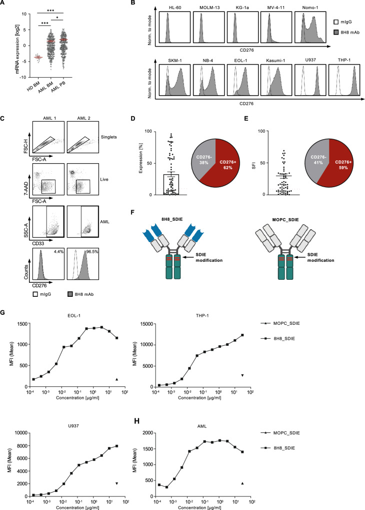
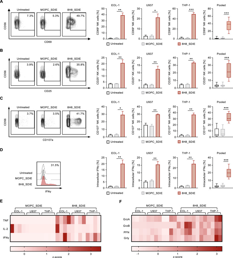
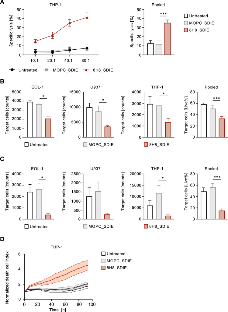
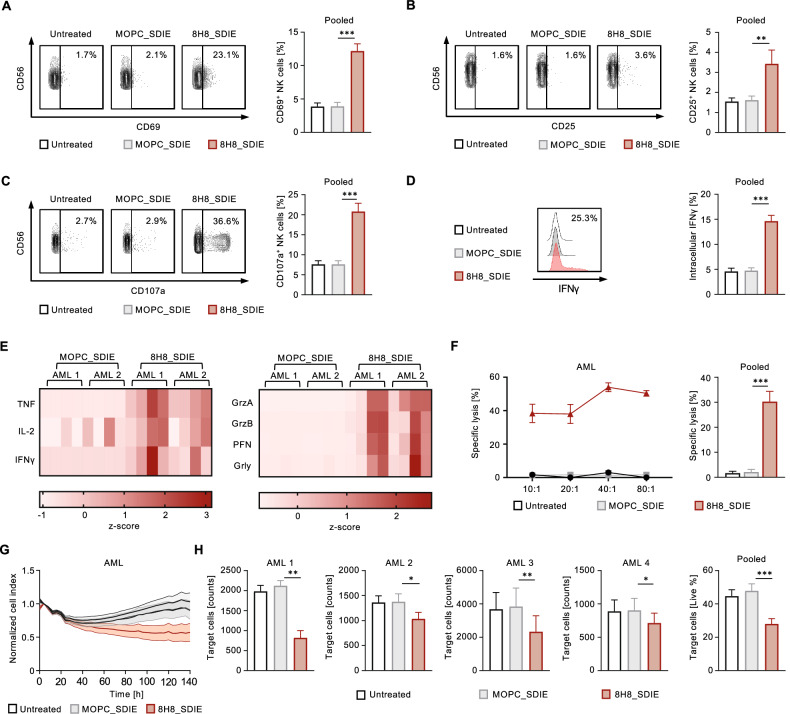
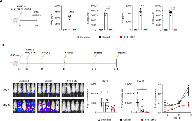
Acute myeloid leukemia (AML) remains a therapeutic challenge despite recent therapeutic advances. Although monoclonal antibodies (mAbs) engaging natural killer (NK) cells via antibody-dependent cellular cytotoxicity (ADCC) hold promise in cancer therapy, almost none have received clinical approval for AML, so far. Recently, CD276 (B7-H3) has emerged as a promising target for AML immunotherapy, due to its high expression on leukemic blasts of AML patients. Here, we present the preclinical development of the Fc-optimized CD276 mAb 8H8_SDIE with enhanced CD16 affinity. We demonstrate that 8H8_SDIE specifically binds to CD276 on AML cell lines and primary AML cells and induces pronounced NK cell activation and degranulation as measured by CD69, CD25, and CD107a. Secretion of IFNγ, TNF, granzyme B, granulysin, and perforin, which mediate NK cell effector functions, was induced by 8H8_SDIE. A pronounced target cell-restricted lysis of AML cell lines and primary AML cells was observed in cytotoxicity assays using 8H8_SDIE. Finally, xenograft models with 8H8_SDIE did not cause off-target immune activation and effectively inhibited leukemia growth in vivo. We here present a novel attractive immunotherapeutic compound that potently induces anti-leukemic NK cell reactivity in vitro and in vivo as treatment option for AML.
© 2024. The Author(s).
Conflict of interest statement
The authors declare no competing interests.
Figures
Similar articles
-
Preclinical Evaluation of a B7-H3 Targeting Antibody Enhancing NK Cell-Mediated Cytotoxicity for Ovarian Cancer Treatment.Immunotargets Ther. 2025 Jul 11;14:735-753. doi: 10.2147/ITT.S521008. eCollection 2025. Immunotargets Ther. 2025. PMID: 40666622 Free PMC article.
-
B7-H3-targeting Fc-optimized antibody for induction of NK cell reactivity against sarcoma.Front Immunol. 2022 Oct 7;13:1002898. doi: 10.3389/fimmu.2022.1002898. eCollection 2022. Front Immunol. 2022. PMID: 36275693 Free PMC article.
-
An Fc-optimized CD133 antibody for induction of NK cell reactivity against myeloid leukemia.Leukemia. 2017 Feb;31(2):459-469. doi: 10.1038/leu.2016.194. Epub 2016 Jul 20. Leukemia. 2017. PMID: 27435001
-
Acute myeloid leukemia and novel biological treatments: monoclonal antibodies and cell-based gene-modified immune effectors.Immunol Lett. 2013 Sep-Oct;155(1-2):43-6. doi: 10.1016/j.imlet.2013.09.013. Epub 2013 Sep 25. Immunol Lett. 2013. PMID: 24076117 Review.
-
B7-H3 in acute myeloid leukemia: From prognostic biomarker to immunotherapeutic target.Chin Med J (Engl). 2024 Nov 5;137(21):2540-2551. doi: 10.1097/CM9.0000000000003099. Epub 2024 Apr 9. Chin Med J (Engl). 2024. PMID: 38595093 Free PMC article. Review.
Cited by
-
Tumor Immunotherapy Targeting B7-H3: From Mechanisms to Clinical Applications.Immunotargets Ther. 2025 Mar 27;14:291-320. doi: 10.2147/ITT.S507522. eCollection 2025. Immunotargets Ther. 2025. PMID: 40171330 Free PMC article. Review.
-
NK cell-based immunotherapy strategies for myeloid leukemia.Front Immunol. 2025 Jul 14;16:1621885. doi: 10.3389/fimmu.2025.1621885. eCollection 2025. Front Immunol. 2025. PMID: 40726987 Free PMC article. Review.
-
Expression and Prognostic Value of a Novel B7-H3 (CD276) Antibody in Acute Myeloid Leukemia.Cancers (Basel). 2024 Jul 4;16(13):2455. doi: 10.3390/cancers16132455. Cancers (Basel). 2024. PMID: 39001517 Free PMC article.
-
Research progress of B7-H3 in malignant tumors.Front Immunol. 2025 May 30;16:1586759. doi: 10.3389/fimmu.2025.1586759. eCollection 2025. Front Immunol. 2025. PMID: 40519928 Free PMC article. Review.
-
Preclinical Evaluation of a B7-H3 Targeting Antibody Enhancing NK Cell-Mediated Cytotoxicity for Ovarian Cancer Treatment.Immunotargets Ther. 2025 Jul 11;14:735-753. doi: 10.2147/ITT.S521008. eCollection 2025. Immunotargets Ther. 2025. PMID: 40666622 Free PMC article.
References
Publication types
MeSH terms
Substances
Grants and funding
- MA 9302/2-1/Deutsche Forschungsgemeinschaft (German Research Foundation)
- MA 9302/2-1/Deutsche Forschungsgemeinschaft (German Research Foundation)
- MA 9302/2-1/Deutsche Forschungsgemeinschaft (German Research Foundation)
- MA 9302/2-1/Deutsche Forschungsgemeinschaft (German Research Foundation)
- MA 9302/2-1/Deutsche Forschungsgemeinschaft (German Research Foundation)
- MA 9302/2-1/Deutsche Forschungsgemeinschaft (German Research Foundation)
- MA 9302/2-1/Deutsche Forschungsgemeinschaft (German Research Foundation)
- MA 9302/2-1/Deutsche Forschungsgemeinschaft (German Research Foundation)
- MA 9302/2-1/Deutsche Forschungsgemeinschaft (German Research Foundation)
- 70113496/Deutsche Krebshilfe (German Cancer Aid)
- 70113496/Deutsche Krebshilfe (German Cancer Aid)
- 70113496/Deutsche Krebshilfe (German Cancer Aid)
- 70113496/Deutsche Krebshilfe (German Cancer Aid)
- 70113496/Deutsche Krebshilfe (German Cancer Aid)
- 70113496/Deutsche Krebshilfe (German Cancer Aid)
- 70113496/Deutsche Krebshilfe (German Cancer Aid)
- 70113496/Deutsche Krebshilfe (German Cancer Aid)
- 70113496/Deutsche Krebshilfe (German Cancer Aid)
LinkOut - more resources
Full Text Sources
Medical
Research Materials
