. 2024 Apr 18;22(1):370.
doi: 10.1186/s12967-024-05144-4.

Discovery of a novel and highly selective JAK3 inhibitor as a potent hair growth promoter

Affiliations

Discovery of a novel and highly selective JAK3 inhibitor as a potent hair growth promoter

Md Mehedi Hossain et al. J Transl Med. .

Abstract

JAK-STAT signalling pathway inhibitors have emerged as promising therapeutic agents for the treatment of hair loss. Among different JAK isoforms, JAK3 has become an ideal target for drug discovery because it only regulates a narrow spectrum of γc cytokines. Here, we report the discovery of MJ04, a novel and highly selective 3-pyrimidinylazaindole based JAK3 inhibitor, as a potential hair growth promoter with an IC50 of 2.03 nM. During in vivo efficacy assays, topical application of MJ04 on DHT-challenged AGA and athymic nude mice resulted in early onset of hair regrowth. Furthermore, MJ04 significantly promoted the growth of human hair follicles under ex-vivo conditions. MJ04 exhibited a reasonably good pharmacokinetic profile and demonstrated a favourable safety profile under in vivo and in vitro conditions. Taken together, we report MJ04 as a highly potent and selective JAK3 inhibitor that exhibits overall properties suitable for topical drug development and advancement to human clinical trials.

Keywords: 3-pyrimidinylazaindole; Alopecia areata; Baricitinib; Hair follicles; JAK-STAT signalling; Ritlecitinib; Tofacitinib.


Conflict of interest statement

The authors declare that they have no competing interests.

Figures

Fig. 1
Fig. 1
Identification of MJ04 as JAK3 inhibitor and its biological characteristics. A The chemical structure of compound MJ04. B Inhibitory activity and selectivity of MJ04 for JAK family kinases at Km and 1 mM ATP concentration. C The IC50 of MJ04 and tofacitinib inhibiting JAK3 at Km ATP concentration. D The inhibitory potency and selectivity of MJ04 against a panel of 10 kinases at Km ATP concentration. E Intermolecular interactions of MJ04 with JAK1 (3EYG), JAK2 (6TPD) and JAK3 (5WFJ) kinase domain: E (a) Multiple sequence alignment of JAK1, JAK2 and JAK3 with conserved regions labelled as G loop (glycine rich loop, L881-E890-JAK1, L855-E864-JAK2, L828-E837-JAK3), hinge region (M956-L968-JAK1, M929-L941-JAK2, M902-L914-JAK3) catalytic loop (H1001-L1010-JAK1, H974-L981-JAK2, H947-L956-JAK3) and A loop (activation loop, D1021-E1051-JAK1, D994-E1024-JAK2, D967E997-JAK3) indicating the conserved residues in each region. E (b) represents the superimposed structure of JAK1, JAK2 and JAK3 with inhibitor molecule MJ04 present in the hinge region and different regions are coloured and labelled as hinge region (hot pink), catalytic loop (orange), activation loop (blue) and glycine rich loop (cyan), the E (c), E (d) and E (e) is representing the enlarged view of intermolecular interactions of MJ04 with JAK1, JAK2 and JAK3 respectively with inhibitor MJ04 in stick coloured in red and heteroatoms coloured differently, hydrophobic interacting residues in wire and magenta colour and hydrogen bonding residues in stick coloured in yellow with heteroatoms differently coloured, H-bonds are represented as red coloured dotted lines with length labelled
Fig. 2
Fig. 2
Selective Inhibition of the JAK3-Dependent Signaling Pathway by MJ04. A Assessment of cell cytotoxicity induced by MJ04 and its determination of IC50 values across different cancer cell lines. B Examination of the impact of MJ04 on the phosphorylation status of JAK1, JAK3, and STAT3. C Evaluation of the effect of Tofacitinib on the phosphorylation of JAK1, JAK3, and STAT3. D Assessment of MJ04’s impact on STAT3 phosphorylation in the presence of IL-6 of A549. GAPDH served as the loading control. Densitometry analysis results for each immunoblot are depicted in the accompanying graph to the right. All data analysis was performed using MS Excel, GraphPad Prism-V8.0 Software, and Image-J. Statistical significance was determined through one-way analysis of variance (ANOVA). Data points are presented as mean ± SD, and significance values are denoted as *P < 0.05, **P < 0.005, ***P < 0.0005, and ****P < 0.00005."
Fig. 3
Fig. 3
Effect of MJ04 on hair regrowth in the DHT induced AGA mice model. The shaved back skin of C57BL/6J mice were daily treated with 0.5% testosterone for 1 h prior to topical application of various concentration of tofacitinib, MJ04 and baricitinib for 28 days. A MJ04 administration induced the telogen-anagen transition. Digital photographs were capture from a representative area using Nikon digital camera (n = 8 mice), B Qualitative analysis:—The quantification of anagen initiation was assessed by observing changes in the dorsal skin colour of C57BL/6 mice, a phenomenon closely linked to the onset of anagen phase. Anagen quantification was carried out by employing threshold analysis on dorsal skin colouration using ImageJ. The darkening of the skin, attributed to melangenesis, strongly correlates with progression of anagen. This analysis is visually depicted as a heatmap. Here a = Control; b = Testosterone treated; c = Tofacitinib treated (0.08mg/kg), d = Baricitinib treated (0.04mg/kg), e = MJ04 treated (0.04mg/kg) and f = MJ04 treated (0.08mg/kg), C Dorsal skin samples from C57BL/6J mice were shaved and collected for histological and molecular analyses on Days 7, 14, 21, and 28. The histopathological examination of the samples were executed. n = 8 mice per group H&E stained skin section of C57BL/6J mice (× 200 magnification). a. Normal histoarchitecture with hair follicles. b. Epidermal thinning formula image with very tiny hair follicles formula image. c. Minimal epidermal thinning with few hair follicles on Days 7 and 14 and increased hair follicles on Days 21 and 28 formula image. d. Minimal degree of epidermal thickening with increased distinct hair follicles formula image in the dermal and hypodermal regions. e. Minimal degree of epidermal atrophy formula image with hair follicles on Days 7 and 14 and minimal epidermal thickening formula image with increased hair follicles formula image on Days 21 and 28. f. Normal epidermis with increased hair follicles formula image 
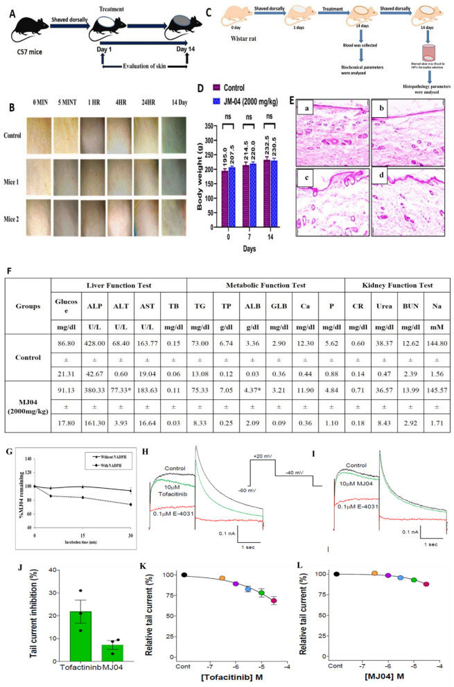
on Days 14 and 28. D Mean Weekly Body Weight: Monitoring of mean weekly body weight of DHT induced AGA mice model throughout the evaluation of MJ04’s impact on hair regrowth. Here, the groups are denoted as follows a = Control; b = Testosterone treated; c = Tofacitinib treated (0.08mg/kg), d = Baricitinib treated (0.04mg/kg), e = MJ04 treated (0.04mg/kg) and f = MJ04 treated (0.08mg/kg). The body weight of the induced AGA mice model was recorded on days 1, 7, 14, 21, and 28
Fig. 4
Fig. 4
MJ04 induces early onset in anagen development, hair cycle progression in nude mice, and growth of human hair follicles under ex-vivo conditions. A The treatment with MJ04 resulted in the transition from telogen-anagen, as evidenced by the transformation of initially hairless dorsal skin to white-haired skin. Digital photographs were captured from the representative areas using a Nikon digital camera (n = 5 mice). a- Control, b-Vehicle control, cTofacitinib (0.8mg/Kg), d-Tofacitinib (0.08mg/kg). e-MJ04 (0.08mg/Kg), f- MJ04 (0.04mg/kg), g- MJ04 (0.016mg/kg), h- Baricitinib (0.1mg/kg), i- Baricitinib (0.04 mg/kg), j- Baricitinib- (0.02 mg/Kg). B Mean Weekly Body Weight: Monitoring of mean weekly body weight of athymic nude mice (NU/J Foxn1nu) mice model throughout the evaluation of MJ04's impact on hair regrowth. Here a- Control, b-Vehicle control, c-Tofacitinib (0.8mg/Kg), d-Tofacitinib (0.08mg/kg). e-MJ04 (0.08mg/Kg), f- MJ04 (0.04mg/kg), g- MJ04 (0.016mg/kg), h- Baricitinib (0.1mg/kg), i- Baricitinib (0.04 mg/kg), j- Baricitinib- (0.02 mg/Kg). The body weight of the nude mice was recorded on days 1, 7, 14, 21, and 28. C The skin samples collected from athymic nude mice (NU/J Foxn1nu) were preserved in 10% neutral buffered formalin and subsequently subjected to tissue sectioning. Skin sections (10 µm) of nude mice were stained with haematoxylin and eosin (H&E) and viewed under 10× and 40× objectives of the light microscope (Magnus, Mx21i). The histopathological examination of the samples was performed, and the hair follicles in the skin sections were graded on a scale ranging from 0 to 4 (++++). H&E stained the skin section of nude mice (×100 magnification). a and b (control & vehicle control): Normal histoarchitecture with the intact epidermis (formula image, dermis formula image, hypodermis formula image, panniculus carnosus, and adventitia formula image. c and d (Tofacitinib citrate—0.08 and 0.8 mg/kg): Mild epidermal thickening with the increased number of hair follicles [formula image] (++++). e (MJ04—0.08 mg/kg): Minimal epidermal thickening with moderate hair follicle (++). f (MJ04—0.04 mg/kg): Mild epidermal thinning with increased hair follicles in the dermis and hypodermis (++++). g (MJ04—0.016 mg/kg): Mild epidermal thickening with increased hair follicles (+++). h (Baricitinib–0.1 mg/kg): Increased and morphologically distinct hair follicles formula image in the dermis and hypodermis regions (+++). i and j (Baricitinib—0.04 and 0.02 mg/kg): Moderate degree of epidermal thickening formula image with more hair follicles in the dermis and hypodermis regions (++++), and D MJ04 promotes the growth of human hair follicles under ex-vivo conditions. (a) Control group: (i) Basal length, (ii) Length posttreatment, (b) Experimental group treated with 100 μl of MJ04 (0.50 μg/μl): (i) Basal length, (ii) Length post-experiment, and (c) Experimental group treated with 100 μl of MJ04 (0.25 μg/μl): (i) Basal length, (ii) Length post-treatment. The red bracket indicates the length of the human hair follicle
Fig. 5
Fig. 5
MJ04 inhibits the differentiation of naïve T-cell into Th1 and Tc1 subsets, also inhibits the level of proinflammatory cytokines in LPS primed macrophages. Splenoctyes from healthy mice were used as a source of T-cells, Spelnocytes were stimulated with anti-CD3/C28 in the presence and absence of MJ04 (0.01, 0.1, 1 and 10 µM) for 96 h and before harvesting these cells were further stimulated with PMA/Ionomycin for additional 4 h. AD is the gating strategy to deduce the cell of interest. The expression of IFN-γ in the CD4+(C, D) and CD8+ (E, F) T-cells were analyzed by flowcytometry. In another set of experiments, mouse macrophage (J774a.1) were induced with LPS and then treated with different concentration of MJ04 and Tofacitinib. The level of pro inflammatory cytokines (TNF-α, IL1-β and IL-6) were measured by sandwich ELISA from LPS induced macrophages (GI). Each data point is representative of n = 3). Data points are represented as mean ± SEM and value of significance represents *P < 0.05, **P < 0.005, ***P < 0.0005 and ****P < 0.00005
Fig. 6
Fig. 6
Preclinical safety and toxicity profile of MJ04. A Experimental Overview: The dorsal skin of C57BL/6J mice was shaved, and subsequent treatments were applied to the dorsal skin surface on the following day. B Acute Dermal Toxicity Assessment: Signal dose acute dermal toxicity tests were conducted in C57BL/6 mice. C Experimental overview of Median Lethal Dose (LD50) Determination: Acute dermal toxicity tests were conducted in Wistar rats. Limited testing included a single dose at 2 g/kg body weight. D Mean Weekly Body Weight: Monitoring of mean weekly body weight during the acute dermal toxicity assessment of MJ04. E Histopathological Analysis of dorsal skin tissue: Haematoxylin and eosin images depict the dorsal skin of mice (n = 5 mice per group). The histopathology images are presented at 10× magnification and show the skin of the control (a and b) and MJ04 treated group (2000 mg/kg) (c and d). F Mean serum biochemistry of acute dermal toxicity of MJ04. The values are expressed as mean ± SD (n = 3); *Significant difference from the control group at P < 0.05; b.w.: Body weight; GLU: Glucose, ALP: Alkaline Phosphatase, ALT: Alanine Transaminase, AST: Aspartate Amino Transferase, TB: Total Bilirubin, TG: Triglycerides, TP: Total Protein, ALB: Albumin, GLB: Globulin, CR: Creatinine, BUN: Blood Urea Nitrogen, Ca: Calcium, P: Phosphorus, Na: Sodium; No significant difference in the means of treatment groups and control group at 0.05 level. G Represents the metabolic stability of MJ04 in human liver microsomes. HL shows the effect of MJ04 on hERG channel currents. Representative current traces show the effect of control (0.1% DMSO, black traces), 0.1 µM E-4031 (red traces), 10 µM Tofacitinib (green traces, H), 10 µM MJ04 (green traces, I). The recording protocol employed is shown in inset. J Mean tail current % inhibition for each drug is plotted. K, and L shows the summary dose response for 5 concentrations of the Tofacitinib (K) and MJ04 (L) ranging from 0.3 µm to 30 µM. Data points are the mean ± SEM (n = 3)
Fig. 7
Fig. 7
The model shows the fate of hair follicles under alopecia pathogenesis and restoration of growth of hair follicles upon blocking the production of pro-inflammatory cytokines with MJ04 as JAK3 inhibitor. A Shows the healthy hair follicle in the anagen phase of the growth cycle. B Shows the disrupted microenvironment of hair follicles due to infiltration of proinflammatory cytokines and immune cells during alopecia pathogenesis. C Blocking JAK-STAT signaling in immune cells inhibits the production of pro-inflammatory cytokines that restore back homeostasis around hair follicles allowing them to enter into a new growth cycle

References

    1. Cotsarelis G, Millar SE. Towards a molecular understanding of hair loss and its treatment. Trends Mol Med. 2001;7:293–301. doi: 10.1016/S1471-4914(01)02027-5. - DOI - PubMed
    1. Daszczuk P, Mazurek P, Pieczonka TD, Olczak A, Boryń ŁM, Kobielak KJ. An intrinsic oscillation of gene networks inside hair follicle stem cells: an additional layer that can modulate hair stem cell activities. Front Cell Dev Biol. 2020;8:595178. doi: 10.3389/fcell.2020.595178. - DOI - PMC - PubMed
    1. Sadick NS, Callender VD, Kircik LH, Kogan SJ. New insight into the pathophysiology of hair loss trigger a paradigm shift in the treatment approach. J Drugs Dermatol. 2017;16:s135–s140. - PubMed
    1. Williams R, Pawlus AD, Thornton MJJ. Getting under the skin of hair aging: the impact of the hair follicle environment. Exp Dermatol. 2020;29:588–597. doi: 10.1111/exd.14109. - DOI - PubMed
    1. Weger N, Schlake TJ. Igf-I signalling controls the hair growth cycle and the differentiation of hair shafts. J Investig Dermatol. 2005;125:873–882. doi: 10.1111/j.0022-202X.2005.23946.x. - DOI - PubMed

Publication types

LinkOut - more resources