. 2024 Apr 4:15:1354617.
doi: 10.3389/fimmu.2024.1354617. eCollection 2024.

Enrichment of type 1 innate lymphoid cells in the course of human atherosclerotic plaque development suggests contribution to atherogenesis

Affiliations

Enrichment of type 1 innate lymphoid cells in the course of human atherosclerotic plaque development suggests contribution to atherogenesis

Kartika R Pertiwi et al. Front Immunol. .

Abstract

Introduction: Innate lymphoid cells (ILCs) have been implicated in multiple pathologic conditions, including atherogenesis, as documented in experimental mice studies, however, their role in atherosclerosis in humans remains unexplored.

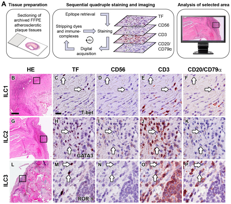
Methods: Here, we identify ILCs and their dynamics in early, advanced, and complicated human carotid- and aortic atherosclerotic plaques, using a multiplex immunohistochemical quadruple-staining technique with prototypic transcription factors T-bet, GATA3, or RORgt for identification of the ILC1, ILC2 and ILC3 subsets, respectively, in combination with lineage markers CD3, CD20/ CD79a and CD56 to exclude other lymphoid cell types. ILC subsets were quantified, and to put this in perspective, their numbers were expressed as percentage of the total number of infiltrated lymphoid cells and related to the frequency of conventional T cells, B cells, NK cells, and NKT cells.

Results: All ILC subsets were present in every different stage of atherogenesis. ILC1s were the most abundant ILC subset, and their numbers significantly increased in the course of plaque development, but paradoxically, their relative frequency was reduced because of a higher increment of T cells and B cells. The numbers of ILC2s and ILC3s also gradually increased, but this trend did not achieve significance. T cell subsets always significantly outnumbered their ILC counterparts, except for the early lesions where the proportion of ILC1s was markedly higher, albeit not significant.

Discussion: The high abundance of ILC1s in the early stages and further significant enrichment in later stages, suggest they may participate in the initiation and development of atherogenesis, and thus, may represent a novel target to prevent or treat atherosclerosis.

Keywords: atherosclerosis; immunohistochemistry; inflammation; innate immunity; innate lymphoid cells; lymphocytes.


Conflict of interest statement

The authors declare that the research was conducted in the absence of any commercial or financial relationships that could be construed as a potential conflict of interest.

Figures

Figure 1
Figure 1
Identification of ILC1s, ILC2s and ILC3s in the intima of human atherosclerotic lesions. (A) Workflow of quadruple staining. Hematoxylin-eosin (HE) overview (B, G, L) and detailed (immunohistochemical) staining of carotid atherosclerotic plaques showing the identification of ILC1s (C–F), ILC2s (H–K) and ILC3s (M–P). Sections were stained with either T-bet (C), GATA3 (H) or RORγt (M), and subsequently digitized and destained. This workflow was repeated for CD56 (D, I, N), CD3 (E, J, O) and CD20/CD79α (F, K, P). ILCs were identified by positive staining with the transcription factor (TF), but being negative for lineage markers CD56, CD3 and CD20/CD79α. Arrows indicate ILCs in sequentially stained sections. Scale bar in HE overview (B): 200 µm. Scale bar in high power detail (C): 25 µm. TF, transcription factor.
Figure 2
Figure 2
Quantification of ILC1s, ILC2s and ILC3s in different stages of atherosclerotic plaque development. (A) Numbers of ILC subsets (cells/mm2) present in the different stages (early, advanced, or complicated) of atherosclerotic plaques. (B) Proportion of ILC1s, ILC2s and ILC3s present in each plaque category, being expressed as a percentage of the total number of lymphoid cells. Data are expressed as box and whisker plots, with the median indicated by a bold horizontal line and the mean indicated by a + symbol. ILC2 and ILC3 are depicted in black, blue, and red, respectively, whereas the different stages are indicated by dots (early), diamonds (advanced), and triangles (complicated). (*p < 0.05).
Figure 3
Figure 3
Identification of ILC1s in an early atherosclerotic plaque (fatty streak). (A) Hematoxylin-eosin staining. (B) False color multiplex staining showing T-bet in cyan and CD3 in green (closed and open arrowheads, respectively). This section was also stained for NK cells (CD56) and B cells (CD20/CD79α), but these cells were not present in this high power field. (C) Immunohistochemical double staining for T-bet (in blue, open arrowhead) and CD68 (red, closed arrowhead) showing that intralesional macrophages do not express T-bet. Scale bar equals 100 µm in (A, B) and 50 µm in (C). i, intima; m, media.
Figure 4
Figure 4
Identification of T1, T2 and T17 cells in the intima of human atherosclerotic lesions. Hematoxylin-eosin (HE) overview (A, F, K) and detailed staining of carotid atherosclerotic plaques showing the identification of T1 (B–E), T2 (G–J) and T17 (L–O) cells. Sections were stained with either T-bet (B), GATA3 (G) or RORγt (L), and subsequently digitized and destained. This procedure was repeated for CD56 (C, H, M), CD3 (D, I, N) and CD20/CD79α (E, J, O). T1, T2 and T17 cells were identified by positive staining with TF and CD3, but being negative for CD56 and CD20/CD79α. Arrows indicate transcription factor (TF) positive T cells. Scale bar in HE overview (A): 200 µm. Scale bar in high power detail (B): 25 µm.
Figure 5
Figure 5
Quantification of T1, T2 and T17 cells in different stages of atherosclerotic plaque development. (A) Numbers of T1, T2 and T17 cells (cells/mm2) present in the different stages (early, advanced, or complicated) of atherosclerotic plaques. (B) Proportion of T1, T2 and T17 cells present in each plaque category, being expressed as a percentage of the total number of lymphoid cells. Data are expressed as box and whisker plots, with the median indicated by a bold horizontal line and the mean indicated by a + symbol. T1, T2 and T17 cells are depicted in black, blue, and red, respectively, whereas the different stages are indicated by dots (early), diamonds (advanced), and triangles (complicated). (*p < 0.05).
Figure 6
Figure 6
Identification of NK cells, NKT cells and B cells in human atherosclerotic lesions. Sections of carotid atherosclerotic plaques were sequentially stained with T-bet, CD56, CD3, and CD20/CD79α. (A–D) NK cells were identified as CD3CD56+CD20/CD79α cells (arrow). (E–H) NKT cells were identified as CD3+CD56+CD20/CD79α cells (arrow). (I–L) B cells were identified as CD3CD56CD20/CD79α+ cells. Scale bar in (A): 50 µm.
Figure 7
Figure 7
Quantification of NK, NKT and B cells in human atherosclerotic plaques. (A) Number of CD3CD56+CD20/CD79α NK cells, (B) number of CD3+CD56+CD20/CD79α NKT cells, and (C) CD3CD56CD20/CD79α+ B cells present in early, advanced, or complicated atherosclerotic plaques. Data are expressed as box and whisker plots, with the median indicated by a bold horizontal line and the mean indicated by a + symbol. Numbers are expressed as cells/mm2 (*p < 0.05).
Figure 8
Figure 8
Frequencies of ILCs and other types of lymphoid cells in different stages of atherosclerosis. The proportion (means) of different types of innate and adaptive lymphocytes present in early, advanced, or complicated atherosclerotic plaques. The fragmented line indicates the total proportion of innate lymphocytes.

References

    1. Gisterå A, Hansson GK. The immunology of atherosclerosis. Nat Rev Nephrol. (2017) 13:368–80. doi: 10.1038/nrneph.2017.51 - DOI - PubMed
    1. Roy P, Orecchioni M, Ley K. How the immune system shapes atherosclerosis: roles of innate and adaptive immunity. Nat Rev Immunol. (2022) 22:251–65. doi: 10.1038/s41577-021-00584-1 - DOI - PMC - PubMed
    1. Moriya J. Critical roles of inflammation in atherosclerosis. J Cardiol. (2019) 73:22–7. doi: 10.1016/j.jjcc.2018.05.010 - DOI - PubMed
    1. Hansson GK, Hermansson A. The immune system in atherosclerosis. Nat Immunol. (2011) 12:204–12. doi: 10.1038/ni.2001 - DOI - PubMed
    1. de Boer OJ, Becker AE, van der Wal AC. T lymphocytes in atherogenesis-functional aspects and antigenic repertoire. Cardiovasc Res. (2003) 60:78–86. doi: 10.1016/s0008-6363(03)00341-9 - DOI - PubMed