

Proof-of-concept of a robotic-driven photogrammetric scanner for intra-operative knee cartilage repair
- PMID: 38638487
- PMCID: PMC11022211
- DOI: 10.1049/htl2.12054
Proof-of-concept of a robotic-driven photogrammetric scanner for intra-operative knee cartilage repair
Abstract
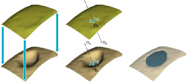
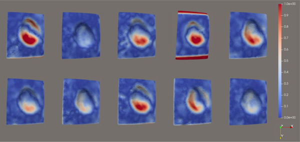
This work presents a proof-of-concept of a robotic-driven intra-operative scanner designed for knee cartilage lesion repair, part of a system for direct in vivo bioprinting. The proposed system is based on a photogrammetric pipeline, which reconstructs the cartilage and lesion surfaces from sets of photographs acquired by a robotic-handled endoscope, and produces 3D grafts for further printing path planning. A validation on a synthetic phantom is presented, showing that, despite the cartilage smooth and featureless surface, the current prototype can accurately reconstruct osteochondral lesions and their surroundings with mean error values of 0.199 ± 0.096 mm but with noticeable concentration on areas with poor lighting or low photographic coverage. The system can also accurately generate grafts for bioprinting, although with a slight tendency to underestimate the actual lesion sizes, producing grafts with coverage errors of -12.2 ± 3.7, -7.9 ± 4.9, and -15.2 ± 3.4% for the medio-lateral, antero-posterior, and craneo-caudal directions, respectively. Improvements in lighting and acquisition for enhancing reconstruction accuracy are planned as future work, as well as integration into a complete bioprinting pipeline and validation with ex vivo phantoms.
Keywords: biological tissues; biomedical imaging; surgery.
© 2023 Vicomtech Foundation. Healthcare Technology Letters published by John Wiley & Sons Ltd on behalf of The Institution of Engineering and Technology.
Conflict of interest statement
The authors declare no conflict of interest.
Figures


Similar articles
-
Collagen 2A Type B Induction after 3D Bioprinting Chondrocytes In Situ into Osteoarthritic Chondral Tibial Lesion.Cartilage. 2021 Dec;13(2_suppl):1755S-1769S. doi: 10.1177/1947603520903788. Epub 2020 Feb 18. Cartilage. 2021. PMID: 32070108 Free PMC article.
-
Robotic-Assisted 3D Bio-printing for Repairing Bone and Cartilage Defects through a Minimally Invasive Approach.Sci Rep. 2019 Mar 6;9(1):3746. doi: 10.1038/s41598-019-38972-2. Sci Rep. 2019. PMID: 30842477 Free PMC article.
-
Accurate 3D reconstruction of bony surfaces using ultrasonic synthetic aperture techniques for robotic knee arthroplasty.Comput Med Imaging Graph. 2017 Jun;58:23-32. doi: 10.1016/j.compmedimag.2017.03.002. Epub 2017 Apr 19. Comput Med Imaging Graph. 2017. PMID: 28448851
-
Three-dimensional Bioprinting for Bone and Cartilage Restoration in Orthopaedic Surgery.J Am Acad Orthop Surg. 2019 Mar 1;27(5):e215-e226. doi: 10.5435/JAAOS-D-17-00632. J Am Acad Orthop Surg. 2019. PMID: 30371527 Review.
-
Autologous tissue transplantations for osteochondral repair.Dan Med J. 2016 Apr;63(4):B5236. Dan Med J. 2016. PMID: 27034191 Review.
References
-
- Krakowski, P. , Karpiński, R. , Jojczuk, M. , Nogalska, A. , Jonak, J. : Knee MRI underestimates the grade of cartilage lesions. Appl. Sci. 11(4), 1552 (2021). 10.3390/app11041552 - DOI
LinkOut - more resources
Full Text Sources
