. 2024 Mar 14;19(1):58-70.
doi: 10.18502/jovr.v19i1.15439. eCollection 2024 Jan-Mar.

Modification of a Selective NTRK2 Agonist and Confirmation of Activity in a Glaucoma-on-a-Chip Model

Affiliations

Modification of a Selective NTRK2 Agonist and Confirmation of Activity in a Glaucoma-on-a-Chip Model

Fatemeh Nafian et al. J Ophthalmic Vis Res. .

Abstract

Purpose: RNYK is a selective agonist of the neurotrophic tyrosine kinase receptor type 2 (NTRK2) which has been screened from a phage-displayed peptide library. Its sequence is SGVYKVAYDWQH, similar to a native NTRK2 ligand, that is, brain-derived neurotrophic factor (BDNF). The current study was performed to recognize and confirm critical residues for RNYK activity in a glaucoma-on-a-chip model.

Methods: We designed a modified RNYK (mRNYK) peptide based on hotspots of the RNYK sequence identified by alanine scanning. The critical residues consisted of tyrosine, valine, aspartic acid, and tryptophan (YVDW); however, lysine and glutamine were also maintained in the final sequence (YKVDWQ) for forming amide bonds and peptide dimerization. The affinity of mRNYK binding was confirmed by testing against NTRK2 receptors on the surface of ATRA-treated SH-SY5Y cells. The neuroprotective effect of mRNYK was also evaluated in cell culture after elevated pressure insult in a glaucoma-on-a-chip model.

Results: The primary amine on the lysine side-chain from one sequence (YKVDWQ) reacted with a γ-carboxamide group of glutamine from the other sequence, forming dimeric mRNYK. In silico, molecular dynamic simulations of the mRNYK-NTRK2 complex showed more stable and stronger interactions as compared to the RNYK-NTRK2 complex. In vitro, mRNYK demonstrated a neuroprotective effect on SH-SY5Y cells under normal and elevated pressure comparable to RNYK. The 50% effective concentration (logEC50) for mRNYK was 0.7009, which was better than RNYK with a logEC50 of 0.8318.

Conclusion: The modified peptide studied herein showed improved stability over the original peptide (RNYK) and demonstrated potential for use as a BDNF agonist with neuroprotective properties for treatment of neurodegenerative disorders such as glaucoma.

Keywords: Brain-derived Neurotrophic Factor; Neuroprotection; Neurotrophic Tyrosine Receptor Kinase; Agonist.


Conflict of interest statement

None.

Figures

Figure 1
Figure 1
Schematic of the EHP set-up to simulate a neurodegenerative condition. A pressure chip was connected to regulators and gauges using pneumatic valves and polyurethane hoses. Adapted from “A lab-on-a-chip model of glaucoma,” by our team at Brain and Behavior, p. 12.[19] Copyright 2020 by Wiley.
Figure 2
Figure 2
Overall binding poses of RNYK and mRNYK peptides. 3D structures of RNYK (a, in blue) and mRNYK (b, in red) were docked with the NTRK2 protein receptor in green (PDB ID 1HCF). Docking results determined hotspot peptide residues for receptor bindings.
Figure 3
Figure 3
Conformational changes in mRNYK-NTRK2 versus RNYK-NTRK2 systems. (a) The plot of RMSD for RNYK (blue) and mRNYK (red) ligands in complex with NTRK2 for 10 ns of simulation. The RMSD of mRNYK shows smaller deviations from its initial position with stable binding interaction. (b) The plot of RMSF for RNYK (blue) versus mRNYK (red) ligands. Most residues of RNYK show more fluctuation with RMSF values above 2.5 Å. The same peak for mRNYK is significantly lower, indicating less motion. (c) The plot of the Rg for mRNYK (red) versus RNYK (blue) in complex with NTRK2. The Rg was nonstop changing for RNYK in complex with NTRK2 during the time of the simulation.
Figure 4
Figure 4
Phase contrast images of SH-SY5Y cells. The scale bars are 50 µm long. (A) ATRA changed the phenotype of cells from N-type to S-type. (B) More ATRA-treated cells are oriented and aligned on the PDL/laminin membrane compared to the naked surface. It showed that PDL/laminin provided an optimum physicochemical environment for neuronal adhesion and expansion.
Figure 5
Figure 5
Normalized relative expression of NTRK2 and p75NTR mRNA from SH-SY5Y treated with either ATRA-BDNF or ethanol in compared to untreated cells (Control). One-way ANOVA analysis revealed a significant overexpression of NTRK2 in treated cells by ATRA-BDNF (4.94 ± 0.08165, gray tracing) compared to its expression in control cells (1 ± 0.04223, black) with adjusted P < 0.0001 (****). In this treatment, the level of p75NTR mRNA was increased a little (1.68 ± 0.01414, gray, ** P = 0.0062). Treated cells by EtOH had a significant overexpression in the p75NTR mRNA (3.58 ± 0.1501) compared to unexposed cells. Although no significant difference was calculated for NTRK2 (P = 0.0815) after EtOH treatment, its mRNA was less compared to its expression level in untreated cells (0.64 ± 0.02438). Adapted from “Peptide selected by phage display increases survival of SH-SY5Y neurons comparable to brain-derived neurotrophic factor,” by our team in Journal of Cellular Biochemistry, p. 7.[7] Copyright 2019 by Wiley.
Figure 6
Figure 6
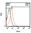
Fluorescence intensity. FITC-conjugated mRNYK bound to the NTRK2 receptors on treated (41.6% FITC+, orang) more than untreated cells (4.80% FITC+, red). In contrast, mRNYK showed no significant binding affinity to induced p75NTR on treated cells (6.12% FITC+, blue).
Figure 7
Figure 7
Time course of EHP-induced cell death. The high pressure was applied to the cells for different times (1–48 hr) followed by 6 hr of recovery. The values represent the percentage of cell death in comparison with control cells not exposed to EHP. The data are the means SD of at least six experiments run in triplicate. *P < 0.05; **P < 0.01 vs control cells not exposed to loading pressure.
Figure 8
Figure 8
Concentration-response curves. Neuroprotective effects of RNYK and mRNYK were compared under an EHP-induced death, (a) and (b) curves, respectively. Different concentrations of peptides were added to the cells, 0.5–5000 ng ml -1 , only before EHP (), during EHP (), during recovery (), or during the entire experiment (). The values represent the percentage of neuroprotection in comparison with control cells (untreated). The best-fit values of LogEC50s for all conditions were 0.7009 for mRNYK and 0.8318 for RNYK (Two-way ANOVA, N = 4, graphs display mean ± SD). Cell survival was evaluated at the end of the recovery period.

References

    1. Soppet D, Escandon E, Maragos J, Middlemas DS, Raid SW, Blair J, et al. The neurotrophic factors brain-derived neurotrophic factor and neurotrophin-3 are ligands for the trkB tyrosine kinase receptor. Cell. 1991;65:895–903. - PubMed
    1. Carter BD, Kaltschmidt C, Kaltschmidt B, Offenhäuser N, Böhm-Matthaei R, Baeuerle PA, et al. Selective activation of NF-κB by nerve growth factor through the neurotrophin receptor p75. Science. 1996;272:542–545. - PubMed
    1. Banfield MJ, Naylor RL, Robertson AG, Allen SJ, Dawbarn D, Brady RL. Specificity in Trk receptor: Neurotrophin interactions: The crystal structure of TrkB-d5 in complex with neurotrophin-4/5. Structure. 2001;9:1191–1199. - PubMed
    1. Kaplan DR, Miller FD. Neurotrophin signal transduction in the nervous system. Curr Opin Neurobiol. 2000;10:381–391. - PubMed
    1. Barker PA. p75NTR is positively promiscuous: Novel partners and new insights. Neuron. 2004;42:529–533. - PubMed

LinkOut - more resources