

Cardiac Tamponade Due to Pericardial Effusion Following Peripherally Inserted Central Catheter: A Single-Institution Case Series
- PMID: 38638757
- PMCID: PMC11025877
- DOI: 10.7759/cureus.56403
Cardiac Tamponade Due to Pericardial Effusion Following Peripherally Inserted Central Catheter: A Single-Institution Case Series
Abstract
Introduction: Although the use of peripherally inserted central catheters (PICCs) has many advantages, misplacement can lead to serious life-threatening complications such as pericardial effusion (PCE) and cardiac tamponade (CT). This report aims to describe four cases of CT resulting from misplaced PICC, which were successfully managed.
Methods: Retrospective analysis of neonates who required PICC insertion and had PCE leading to CT in the Neonatal Intensive Care Unit (NICU) at The Children's Hospital 2, Ho Chi Minh City, Vietnam, during the year 2022.
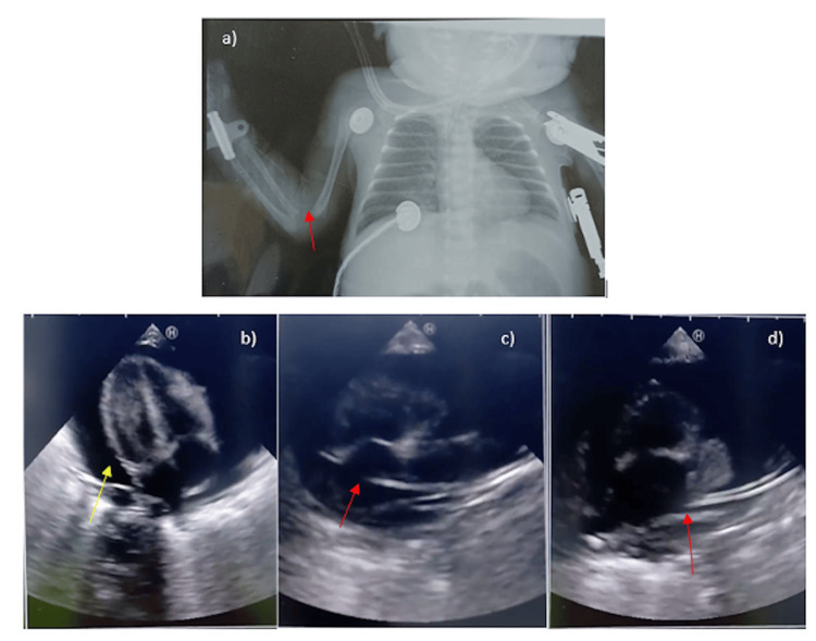
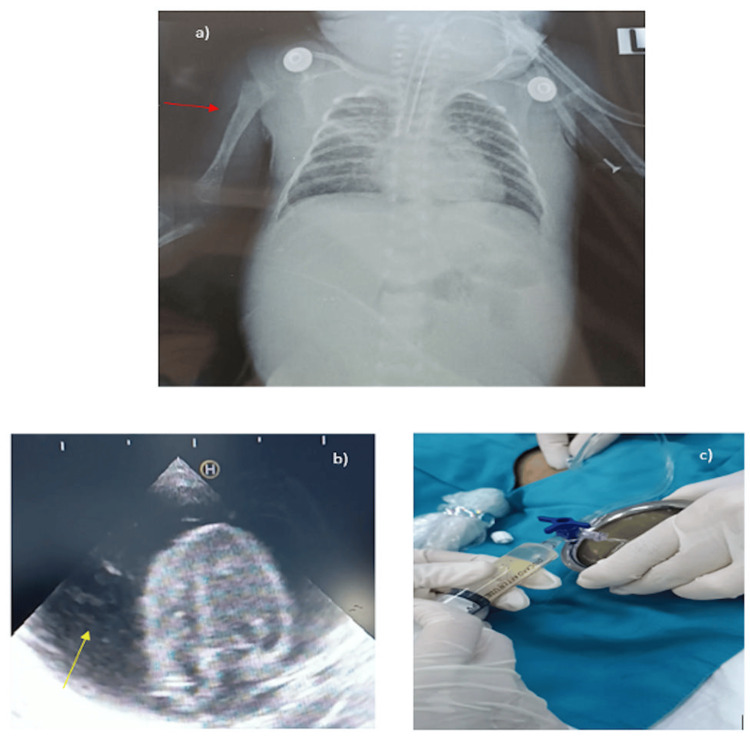
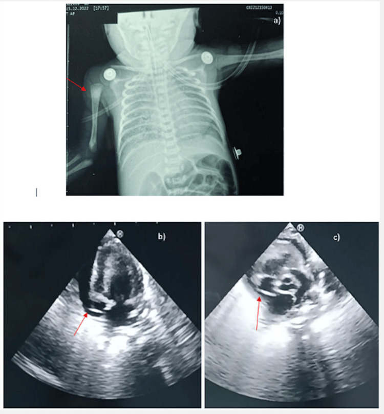
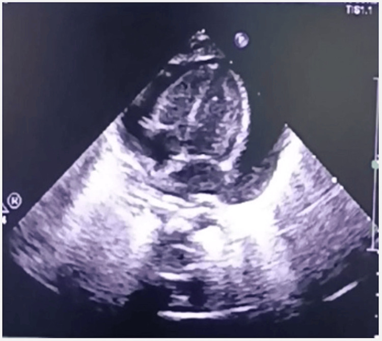
Results: Four cases involved preterm infants at 28-30 weeks gestational age, weighing between 900-1,500 grams. The PCE/CT developed between 3 and 24 days following PICC insertion. The abrupt onset with clinical manifestations that showed hemodynamic instability included sudden deterioration, lethargy, apnea, bradycardia, pale skin, and cardiovascular collapse. We use cardiac point of care ultrasound (POCUS) to assess the condition of these patients and guide the pericardiocentesis procedure. The analysis of the aspirated fluid used for PCE/CT treatment is consistent with the component of parenteral nutrition. No deaths were encountered.
Conclusion: Neonates presenting sudden deterioration following PICC insertion should undergo POCUS to prompt identifying PCE/CT. Timely diagnosis via POCUS, prompt pericardiocentesis, and prevention of misplaced PICC-associated serious complications are crucial. Monitoring of the PICC position twice a week is recommended to avoid life-threatening complications. Additionally, incorporating POCUS for identifying the tip of PICC rather than relying solely on X-ray should be considered in the current protocol.
Keywords: cardiac tamponade; pericardial effusion; peripherally inserted central catheters; point-of-care ultrasound; shock.
Copyright © 2024, Trinh et al.
Conflict of interest statement
The authors have declared that no competing interests exist.
Figures
Similar articles
-
Complications of epicutaneo-caval catheters: Pericardial effusion and cardiac tamponade in three preterm infants.J Vasc Access. 2024 Sep;25(5):1690-1694. doi: 10.1177/11297298231198011. Epub 2023 Sep 20. J Vasc Access. 2024. PMID: 37731340
-
Neonatal cardiac tamponade, a life-threatening complication secondary to peripherally inserted central catheter: a case report.J Med Case Rep. 2022 Jul 28;16(1):305. doi: 10.1186/s13256-022-03506-4. J Med Case Rep. 2022. PMID: 35902974 Free PMC article.
-
Tamponade and massive pleural effusions secondary to peripherally inserted central catheter in neonates-A complication to be aware of.Front Cardiovasc Med. 2023 Feb 17;10:1092814. doi: 10.3389/fcvm.2023.1092814. eCollection 2023. Front Cardiovasc Med. 2023. PMID: 36873398 Free PMC article.
-
Peripherally inserted central catheter related pericardial effusion/cardiac tamponade in neonates: Analysis of two cases and literature review.Medicine (Baltimore). 2023 Oct 27;102(43):e35779. doi: 10.1097/MD.0000000000035779. Medicine (Baltimore). 2023. PMID: 37904403 Free PMC article.
-
Heart in Focus: Advancing Pericardial Effusion Diagnosis With Point-of-Care Ultrasound.Cureus. 2024 Dec 31;16(12):e76681. doi: 10.7759/cureus.76681. eCollection 2024 Dec. Cureus. 2024. PMID: 39886707 Free PMC article. Review.
Cited by
-
Analysis of the preventive effect of upper limb exercise combined with intelligent grip ball on venous thrombosis in patients with peripheral central venous catheters.Am J Transl Res. 2024 Nov 15;16(11):6709-6717. doi: 10.62347/UVLS3384. eCollection 2024. Am J Transl Res. 2024. PMID: 39678600 Free PMC article.
References
-
- Pericardial effusion and tamponade in infants with central catheters. Nowlen TT, Rosenthal GL, Johnson GL, Tom DJ, Vargo TA. Pediatrics. 2002;110:137–142. - PubMed
-
- Pericardial effusion and cardiac tamponade in neonates: sudden unexpected death associated with total parenteral nutrition via central venous catheterization. Warren M, Thompson KS, Popek EJ, Vogel H, Hicks J. https://pubmed.ncbi.nlm.nih.gov/23694791/ Ann Clin Lab Sci. 2013;43:163–171. - PubMed
-
- Cardiac tamponade from peripherally-inserted central venous catheters in neonates: three case reports. Khoo WV, Choo YM, Zahari N, Kamar AA. https://pubmed.ncbi.nlm.nih.gov/34305120/ Med J Malaysia. 2021;76:566–568. - PubMed
Publication types
LinkOut - more resources
Full Text Sources