. 2024 Apr 19:13:e89157.
doi: 10.7554/eLife.89157.

A modified BCG with depletion of enzymes associated with peptidoglycan amidation induces enhanced protection against tuberculosis in mice

Affiliations

A modified BCG with depletion of enzymes associated with peptidoglycan amidation induces enhanced protection against tuberculosis in mice

Moagi Tube Shaku et al. Elife. .

Abstract

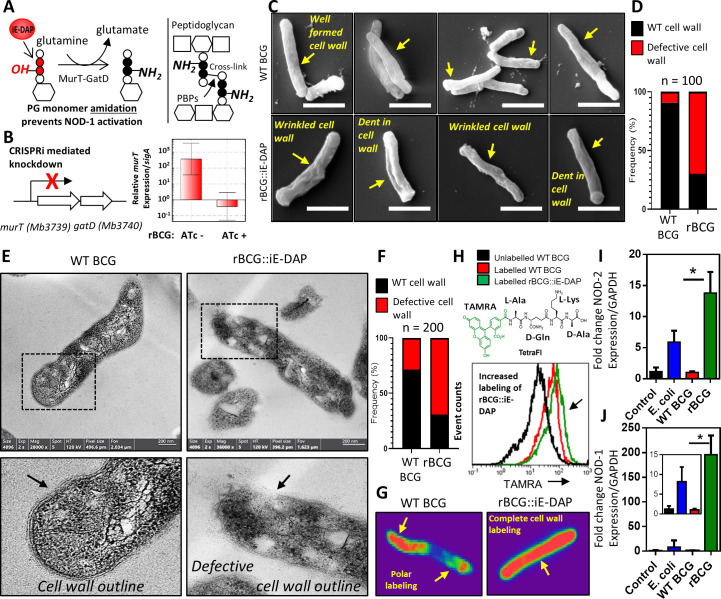
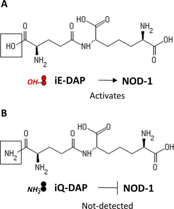
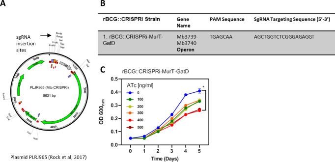
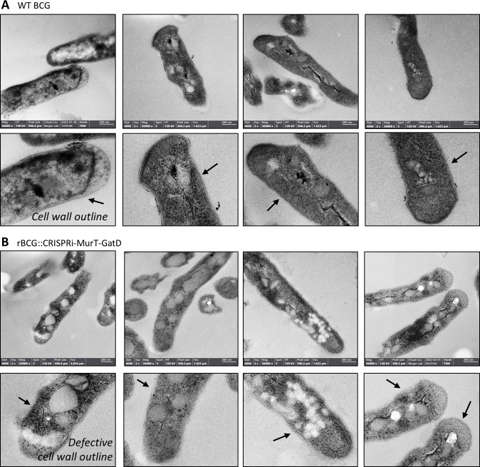
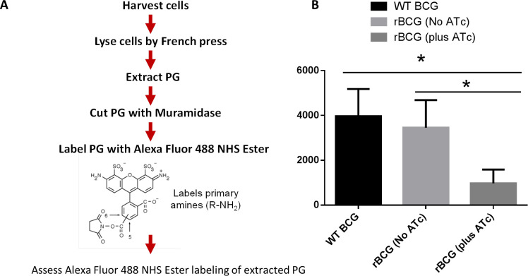
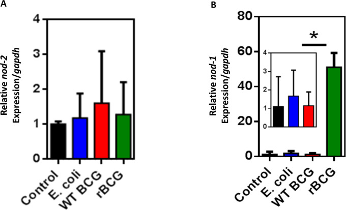
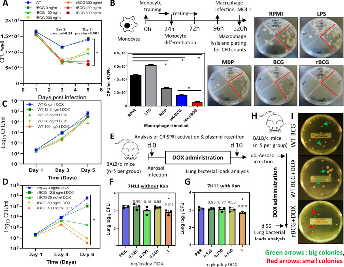
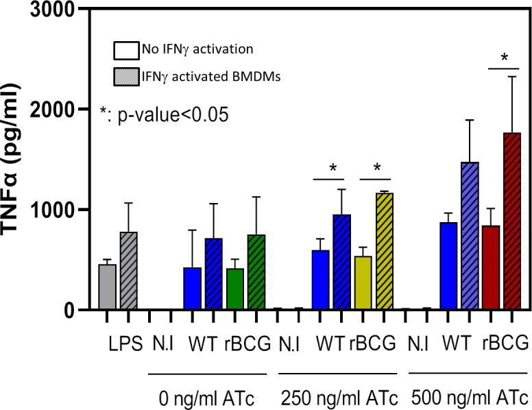
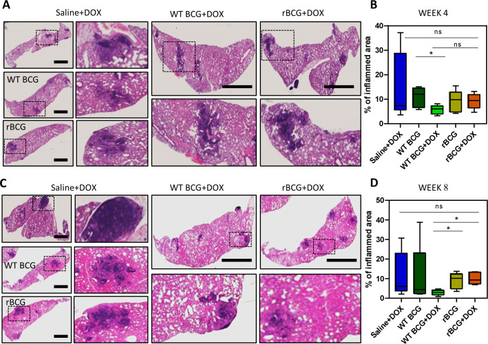
Mechanisms by which Mycobacterium tuberculosis (Mtb) evades pathogen recognition receptor activation during infection may offer insights for the development of improved tuberculosis (TB) vaccines. Whilst Mtb elicits NOD-2 activation through host recognition of its peptidoglycan-derived muramyl dipeptide (MDP), it masks the endogenous NOD-1 ligand through amidation of glutamate at the second position in peptidoglycan side-chains. As the current BCG vaccine is derived from pathogenic mycobacteria, a similar situation prevails. To alleviate this masking ability and to potentially improve efficacy of the BCG vaccine, we used CRISPRi to inhibit expression of the essential enzyme pair, MurT-GatD, implicated in amidation of peptidoglycan side-chains. We demonstrate that depletion of these enzymes results in reduced growth, cell wall defects, increased susceptibility to antibiotics, altered spatial localization of new peptidoglycan and increased NOD-1 expression in macrophages. In cell culture experiments, training of a human monocyte cell line with this recombinant BCG yielded improved control of Mtb growth. In the murine model of TB infection, we demonstrate that depletion of MurT-GatD in BCG, which is expected to unmask the D-glutamate diaminopimelate (iE-DAP) NOD-1 ligand, yields superior prevention of TB disease compared to the standard BCG vaccine. In vitro and in vivo experiments in this study demonstrate the feasibility of gene regulation platforms such as CRISPRi to alter antigen presentation in BCG in a bespoke manner that tunes immunity towards more effective protection against TB disease.

Keywords: NOD1; amidation; infectious disease; microbiology; mouse; mycobacterium; peptidoglycan; recombinant BCG vaccine; tuberculosis.

Plain language summary

Tuberculosis is the leading cause of death from an infectious disease worldwide, partially due to a lack of access to drug treatments in certain countries where the disease is common. The only available tuberculosis vaccine – known as the BCG vaccine – is useful for preventing cases in young children, but is ineffective in teenagers and adults. So, there is a need to develop new vaccines that offer better, and longer lasting, durable protection in people of all ages. During an infection, our immune system recognizes markers known as PAMPs on the surface of bacteria, viruses or other disease-causing pathogens. The recognition of PAMPs by the immune system enables the body to distinguish foreign invading organisms from its own cells and tissues, thus triggering a response that fights the infection. If the body encounters the infectious agent again in the future, the immune system is able to quickly recognize and eliminate it before it can cause disease. Vaccines protect us by mimicking the appearance of the pathogen to trigger the first immune response without causing the illness. The BCG vaccine contains live bacteria that are closely related to the bacterium responsible for tuberculosis called Mycobacterium tuberculosis. Both M. tuberculosis and the live bacteria used in the BCG vaccine are able to hide an important PAMP, known as the NOD-1 ligand, from the immune system, making it harder for the body to detect them. The NOD-1 ligand forms part of the bacterial cell wall and modifying the BCG bacterium so it cannot disguise this PAMP may lead to a new, more effective vaccine. To investigate this possibility, Shaku et al. used a gene editing approach to develop a modified version of the BCG bacterium which is unable to hide its NOD-1 ligand when treated with a specific drug. Immune cells trained with the modified BCG vaccine were more effective at controlling the growth of M. tuberculosis than macrophages trained using the original vaccine. Furthermore, mice vaccinated with the modified BCG vaccine were better able to limit M. tuberculosis growth in their lungs than mice that had received the original vaccine. These findings offer a new candidate vaccine in the fight against tuberculosis. Further studies will be needed to modify the vaccine for use in humans. More broadly, this work demonstrates that gene editing can be used to expose a specific PAMP present in a live vaccine. This may help develop more effective vaccines for other diseases in the future.


Conflict of interest statement

MS, PU, KO, AA, MP, WB No competing interests declared, BK Senior editor, eLife

Figures

Figure 1.
Figure 1.. Phenotypic characterization of rBCG::iE-DAP and NOD-1 activation.
(A) Schematic representation of MurT-GatD mediated PG precursor amidation. (B) murT gene expression measured by quantitative PCR in rBCG::iE-DAP. (C) Scanning electron micrographs of WT BCG (n=45 micrographs, 100 cells counted) and rBCG::iE-DAP (n=48 micrographs, 100 cells counted) grown in media supplemented with 200 ng/ml ATc. Scale bar = 1 µm. (D) Frequency of cells with cell wall defects as seen by SEM. (E) Transmission electron micrographs of WT BCG (n=45 micrographs, 200 cells counted) and rBCG::iE-DAP (n=45 micrographs, 200 cells counted) grown in media supplemented with 200 ng/ml ATc. Scale bar = 200 nm. (F) Frequency of cells with cell wall defects as seen by TEM. (G) MurT-GatD depleted cells labeled with fluorescent BODIPY-FL vancomycin. (H) Flow cytometry analysis of WT BCG and rBCG::iE-DAP cells labelled with a PG amidation reporter probe TAMRA-L-Ala-D-glutamine-L-Lys-D-Ala (TetraFI). (I) nod-2 gene expression measured by quantitative PCR in INFγ activated THP-1 macrophages stimulated with E. coli, WT BCG and rBCG::iE-DAP. (J) nod-1 gene expression measured by quantitative PCR in INFγ activated THP-1 macrophages infected with E. coli, WT BCG and rBCG::iE-DAP. Three independent biological repeats (n=3) were assessed. Student t-test was used for statistical analysis. The error bars represent the standard deviation relative to the mean. *: p-value <0.01.
Figure 1—figure supplement 1.
Figure 1—figure supplement 1.. Molecular structures of iE-DAP and iQ-DAP.
iE-DAP is a NOD-1 ligand and modification of iE-DAP (A) to iQ-DAP (B) leads to evasion of NOD-1 activation.
Figure 1—figure supplement 2.
Figure 1—figure supplement 2.. CRISPRi depletion of MurT-GatD in rBCG::iE-DAP.
(A) Plasmid PLRJ965 encoding dCas9 endonuclease from S. thermophiles and for expression of inserted sgRNA. (B) Table with gene targets and sgRNA targeting sequences. (C) Growth kinetics of rBCG::iE-DAP grown in a range of ATc [0–500 ng/ml]. rBCG::iE-DAP grown media without ATc grows at a similar rate as WT BCG. Activation of the CRISPRi platform in rBCG::iE-DAP with ATc [100–500 ng/ml] resulted in reduced growth in a concentration dependent manner. Three independent biological repeats (n=3) were assessed. Student t-test was used for statistical analysis. The error bars represent the standard deviation relative to the mean. *: p-value <0.01.
Figure 1—figure supplement 3.
Figure 1—figure supplement 3.. TEM reveals defective cell wall of rBCG::iE-DAP.
Transmission electron micrographs of WT BCG (A) and rBCG::iE-DAP (B) grown in media supplemented with 200 ng/ml ATc. Depletion of MurT-GatD causes cell wall defects. Three independent biological repeats were assessed (n=3). Scale bar = 200 nm.
Figure 1—figure supplement 4.
Figure 1—figure supplement 4.. Depletion of MurT and GatD causes reduced PG amidation.
(A) Flow chart representation of the protocol used for assessing PG amidation in MurT-GatD depleted cells by Alexa Fluor 488 NHS Ester labelling of PG in comparison with control cells (ATc- and WT BCG). The Alexa Fluor 488 NHS Ester labels primary amines (R–NH2) also found in PG as a result of amidation. (B). Quantification of Alexa Fluor 488 NHS Ester labeled PG from MurT-GatD depleted cells in comparison to the no ATc control cells. MurT-GatD depletion causes decreased PG amidation which results in decreased labeling with Alexa Fluor 488 NHS Ester. Three independent biological repeats (n=3) were assessed. Student t-test was used for statistical analysis. The error bars represent the standard deviation relative to the mean. *: p-value <0.01.
Figure 1—figure supplement 5.
Figure 1—figure supplement 5.. qPCR of nod-1 and nod-2 expression in non-activated THP-1 macrophages.
(A) nod-2 gene expression measured by quantitative PCR in non-activated THP-1 macrophages stimulated with E. coli, WT BCG and rBCG::iE-DAP. (B) nod-1 gene expression measured by quantitative PCR in non-activated THP-1 macrophages infected with E. coli, WT BCG and rBCG::iE-DAP. Three independent biological repeats (n=3) were assessed. Student t-test was used for statistical analysis. The error bars represent the standard deviation relative to the mean. *: p-value <0.01.
Figure 2.
Figure 2.. Survival of rBCG::iE-DAP in IFNγ activated bone marrow derived macrophages (BMDMs), training of monocytes and activation with doxycycline.
(A) IFNγ-activated BMDMs (1x10⁶ cells) were infected at MOI: 1 with WT BCG and rBCG::iE-DAP. ATc was added to culture media for induction of the CRISPRi system in rBCG::iE-DAP at concentrations ranging from 100 ng/ml – 500 ng/ml and growth of the strains was assessed after 3 and 5 days. (B) Training of U937 monocytes with heat-killed (HK)-rBCG::iE-DAP compared to HK-WT BCG. Shown is also the representative plates for the experiment. (C, D) CFU counts of in vitro grown WT BCG and of rBCG::iE-DAP grown in complete 7H9 medium at varying concentrations of Dox. (E) Determination of the Dox concentration for activation of rBCG::iE-DAP in vivo. Mice were aerosol infected with ~2.5 log10 CFU of rBCG and Dox (0.125–1 mg/kg/day) - was administered by oral gavage for 10 days. (F, G) CFU counts from the experiment shown in panel E. Lung homogenates were plated on both 7H11 with (G) and without (F) kanamycin (25 µg/ml) to assess the loss of the CRISPRi plasmid during in vivo growth. p-values are given above the graphs. (H) Aerosol infection of mice with ~2.5 log10 CFU of WT BCG, rBCG::iE-DAP and administration of Dox (1 mg/kg/day) for 8 weeks. (I) Plates showing the colony size of rBCG::iE-DAP+Dox compared to WT BCG or WT BCG+Dox, recovered from the lungs of aerosol infected mice from the experiment shown in panel H. Three independent biological repeats (n=3) were assessed for the in vitro experiments, the error bars represent the standard deviation relative to the mean. Five mice per group (n=5) were used for the in vivo experiments. Student t-test was used for statistical analysis. The error bars represent the standard deviation relative to the mean. *: p-value <0.05.
Figure 2—figure supplement 1.
Figure 2—figure supplement 1.. Analysis of secreted TNFα levels from non-activated and IFN-activated BMDMs infected with WT BCG and rBCG::iE-DAP at MOI 1:20.
Increased TNF secretion was observed from rBCG::iE-DAP infected IFN-activated BMDMs cultured in media supplemented with 500 ng/ml ATc. LPS was used as a control. Three independent biological repeats (n=3) were assessed. Student t-test was used for statistical analysis. The error bars represent the standard deviation relative to the mean *: p-value <0.01.
Figure 2—figure supplement 2.
Figure 2—figure supplement 2.. Efficacy of 1 mg/kg/day dose of doxycycline for CRISPRi activation.
(A) Schematic representation of mice aerosol infection with WT BCG and rBCG::iE-DAP and analysis of the efficacy of 1 mg/kg/day dose of doxycycline for CRISPRi-MurT-GatD activation in vivo. (B) Day 1 implantation of WT BCG and rBCG::iE-DAP in the lungs of aerosol infected mice. (C) Day 28 bacterial loads of WT BCG and rBCG::iE-DAP aerosol infected mice. (D) PCR amplification of dCas9 in recovered Big (BC) or Small (SC) rBCG::iE-DAP colonies. Lane 1 is 1 kb plus DNA molecular weight marker. (E, F) Growth kinetics of big and small colonies of rBCG::iE-DAP vs WT BCG in liquid broth supplemented with Doxycycline. Three independent biological repeats (n=3) were assessed for the in vitro experiment. Five mice per group (n=5) were used for the in vivo experiments. Statistical analysis was conducted using student t-test. The error bars represent the standard deviation relative to the mean. *: p-value:<0.01.
Figure 3.
Figure 3.. Analysis of rBCG::iE-DAP strain attenuation.
(A) Schematic representation of SCID mice aerosol infection with WT BCG and rBCG::iE-DAP for analysis of strain attenuation. rBCG::iE-DAP activation in vivo was performed by administration of Dox at 1 mg/kg/day. SCID mice (n=5 per group) were aerosol infected with ~2.5 log10 CFU of WT BCG or rBCG::iE-DAP, a WT BCG+Dox group was included as a control. (B) Percent survival of SCID mice following low-dose challenge with WT BCG and rBCG compared to WT BCG+Dox or rBCG+Dox groups. Five mice per group (n=5) were used for the in vivo experiments. student t-test was used for statistical analysis.
Figure 4.
Figure 4.. Efficacy of rBCG::iE-DAP in comparison to standard WT BCG for protection against Mtb H37Rv infection in mice.
(A) Schematic representation of the mouse immunization and Mtb H37Rv challenge protocol. (B) Percentage weight change at week 6 (day 42) immediately prior to Mtb challenge. (C, D) Lung and Spleen bacterial burdens at week 4 and week 8 post-challenge with Mtb. Five mice per group (n=5) were used for the in vivo experiments. Student t-test was used for statistical analysis. The error bars represent the standard deviation relative to the mean. *: p-value <0.05, **: p-value <0.01.
Figure 4—figure supplement 1.
Figure 4—figure supplement 1.. Lumg and spleen weights post-challenge with Mtb.
(A) Lung weights at week 4 and week 8 post-challenge with Mtb. (B) Spleen weights at week 4 and week 8 post-challenge with Mtb. Five mice per group (n=5) were used for the in vivo experiments. Statistical analysis was conducted using student t-test. The error bars represent the standard deviation relative to the mean. *: p-value:<0.01.
Figure 5.
Figure 5.. Histopathological analysis of lung samples.
(A) Histological haematoxylin and eosin (H&E) staining of lung samples at week 4 post Mtb challenge. Scale bar = 2.5 mm. (B) Analysis of percentage of inflamed area (indicated with black boxes) from each mouse lung per immunized group (n=5 per group), shows that rBCG::iE-DAP+Dox immunized mice present with early lung inflammation compared to WT BCG+Dox. (C) H&E staining of lung samples at week 8 post Mtb H37Rv infection. Scale bar = 2.5 mm. (D) Analysis of percentage of inflamed area from each mouse lung (n=5 per group). The percentage inflamed area was evaluated using ImageJ software (NIH) and plotted as whisker box-plots (whiskers represent minimum and maximum values) and a student t-test was used for statistical analysis. Lung sections were derived from 5 mice per group (n=5) from Figure 4a experiments. Statistical analysis was conducted using student t-test. The error bars represent the standard deviation relative to the mean. *: p-value <0.05.

Update of

References

    1. Aldridge BB, Fernandez-Suarez M, Heller D, Ambravaneswaran V, Irimia D, Toner M, Fortune SM. Asymmetry and aging of mycobacterial cells lead to variable growth and antibiotic susceptibility. Science. 2012;335:100–104. doi: 10.1126/science.1216166. - DOI - PMC - PubMed
    1. Angelidou A, Diray-Arce J, Conti MG, Smolen KK, van Haren SD, Dowling DJ, Husson RN, Levy O. BCG as a case study for precision vaccine development: lessons from vaccine heterogeneity, trained immunity, and immune ontogeny. Frontiers in Microbiology. 2020;11:332. doi: 10.3389/fmicb.2020.00332. - DOI - PMC - PubMed
    1. Bekkering S, Blok BA, Joosten LAB, Riksen NP, van Crevel R, Netea MG. In vitro experimental model of trained innate immunity in human primary monocytes. Clinical and Vaccine Immunology. 2016;23:926–933. doi: 10.1128/CVI.00349-16. - DOI - PMC - PubMed
    1. Blok BA, Arts RJW, van Crevel R, Benn CS, Netea MG. Trained innate immunity as underlying mechanism for the long-term, nonspecific effects of vaccines. Journal of Leukocyte Biology. 2015;98:347–356. doi: 10.1189/jlb.5RI0315-096R. - DOI - PubMed
    1. Caruso R, Warner N, Inohara N, Núñez G. NOD1 and NOD2: signaling, host defense, and inflammatory disease. Immunity. 2014;41:898–908. doi: 10.1016/j.immuni.2014.12.010. - DOI - PMC - PubMed

MeSH terms