

GPR1 and CMKLR1 Control Lipid Metabolism to Support the Development of Clear Cell Renal Cell Carcinoma
- PMID: 38640229
- PMCID: PMC11290988
- DOI: 10.1158/0008-5472.CAN-23-2926
GPR1 and CMKLR1 Control Lipid Metabolism to Support the Development of Clear Cell Renal Cell Carcinoma
Abstract
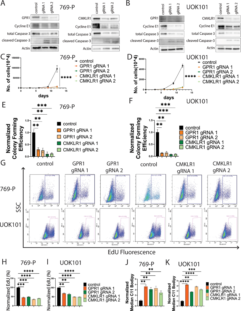
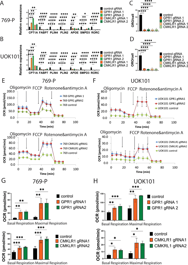
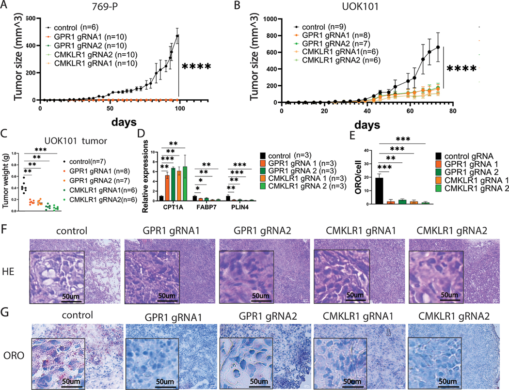
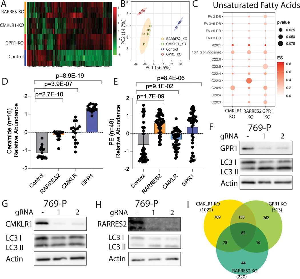
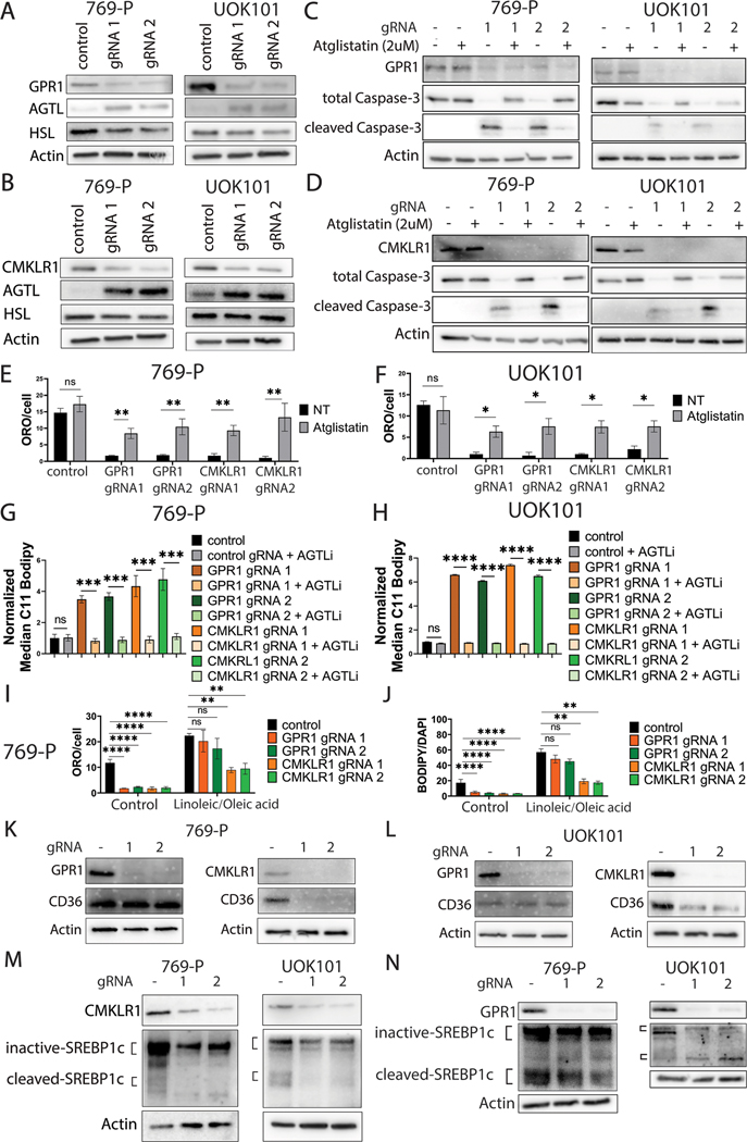
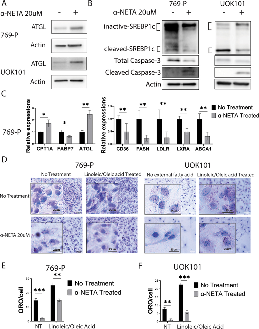
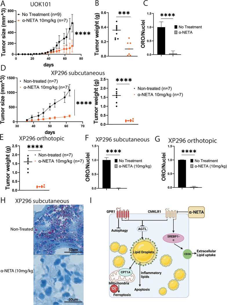
Clear cell renal cell carcinoma (ccRCC), the most common type of kidney cancer, is largely incurable in the metastatic setting. ccRCC is characterized by excessive lipid accumulation that protects cells from stress and promotes tumor growth, suggesting that the underlying regulators of lipid storage could represent potential therapeutic targets. Here, we evaluated the regulatory roles of GPR1 and CMKLR1, two G protein-coupled receptors of the protumorigenic adipokine chemerin that is involved in ccRCC lipid metabolism. Both genetic and pharmacologic suppression of either receptor suppressed lipid formation and induced multiple forms of cell death, including apoptosis, ferroptosis, and autophagy, thereby significantly impeding ccRCC growth in cell lines and patient-derived xenograft models. Comprehensive lipidomic and transcriptomic profiling of receptor competent and depleted cells revealed overlapping and unique signaling of the receptors granting control over triglyceride synthesis, ceramide production, and fatty acid saturation and class production. Mechanistically, both receptors enforced suppression of adipose triglyceride lipase, but each receptor also demonstrated distinct functions, such as the unique ability of CMKLR1 to control lipid uptake through regulation of sterol regulatory element-binding protein 1c and the CD36 scavenger receptor. Treating patient-derived xenograft models with the CMKLR1-targeting small molecule 2-(α-naphthoyl) ethyltrimethylammonium iodide (α-NETA) led to a dramatic reduction in tumor growth, lipid storage, and clear-cell morphology. Together, these findings provide mechanistic insights into lipid regulation in ccRCC and identify a targetable axis at the core of the histologic definition of this tumor that could be exploited therapeutically. Significance: Extracellular control of lipid accumulation via G protein receptor-mediated cell signaling is a metabolic vulnerability in clear cell renal cell carcinoma, which depends on lipid storage to avoid oxidative toxicity.
©2024 American Association for Cancer Research.
Figures
References
-
- Farrukh M, Ali MA, Naveed M, Habib R, Khan H, Kashif T, et al. Efficacy and Safety of Checkpoint Inhibitors in Clear Cell Renal Cell Carcinoma: A Systematic Review of Clinical Trials. Hematology/oncology and stem cell therapy 2023;16:170–85 - PubMed
-
- Rini BI, Campbell SC, Escudier B. Renal cell carcinoma. Lancet 2009;373:1119–32 - PubMed
-
- Barata PC, Rini BI. Treatment of renal cell carcinoma: Current status and future directions. CA Cancer J Clin 2017;67:507–24 - PubMed
-
- Gupta K, Miller JD, Li JZ, Russell MW, Charbonneau C. Epidemiologic and socioeconomic burden of metastatic renal cell carcinoma (mRCC): a literature review. Cancer Treat Rev 2008;34:193–205 - PubMed
MeSH terms
Substances
Grants and funding
- CA253986/National Cancer Institute (NCI)
- R01 CA253986/CA/NCI NIH HHS/United States
- R01 CA254409/CA/NCI NIH HHS/United States
- R35 GM119518/GM/NIGMS NIH HHS/United States
- R01 GM101171/GM/NIGMS NIH HHS/United States
- P50 CA196516/CA/NCI NIH HHS/United States
- P30 CA240139/CA/NCI NIH HHS/United States
- S10 OD012304/OD/NIH HHS/United States
- CA254409/National Cancer Institute (NCI)
- S10OD012304-01/National Cancer Institute (NCI)
- P30CA240139/National Cancer Institute (NCI)
- RP130397/Cancer Prevention and Research Institute of Texas (CPRIT)
- JEK Grant 21K07/Florida Department of Health (DOH)
- P30 CA016672/CA/NCI NIH HHS/United States
- R35GM119518/National Institute of General Medical Sciences (NIGMS)
- GM101171/National Institute of General Medical Sciences (NIGMS)
- U01 CA235510/CA/NCI NIH HHS/United States
LinkOut - more resources
Full Text Sources
Medical
Molecular Biology Databases
