. 2024 Apr 19;10(16):eadl3419.
doi: 10.1126/sciadv.adl3419. Epub 2024 Apr 19.

From 13C-lignin to 13C-mycelium: Agaricus bisporus uses polymeric lignin as a carbon source

Affiliations

From 13C-lignin to 13C-mycelium: Agaricus bisporus uses polymeric lignin as a carbon source

Katharina Duran et al. Sci Adv. .

Abstract

Plant biomass conversion by saprotrophic fungi plays a pivotal role in terrestrial carbon (C) cycling. The general consensus is that fungi metabolize carbohydrates, while lignin is only degraded and mineralized to CO2. Recent research, however, demonstrated fungal conversion of 13C-monoaromatic compounds into proteinogenic amino acids. To unambiguously prove that polymeric lignin is not merely degraded, but also metabolized, carefully isolated 13C-labeled lignin served as substrate for Agaricus bisporus, the world's most consumed mushroom. The fungus formed a dense mycelial network, secreted lignin-active enzymes, depolymerized, and removed lignin. With a lignin carbon use efficiency of 0.14 (g/g) and fungal biomass enrichment in 13C, we demonstrate that A. bisporus assimilated and further metabolized lignin when offered as C-source. Amino acids were high in 13C-enrichment, while fungal-derived carbohydrates, fatty acids, and ergosterol showed traces of 13C. These results hint at lignin conversion via aromatic ring-cleaved intermediates to central metabolites, underlining lignin's metabolic value for fungi.


Figures

Fig. 1.
Fig. 1.. Overview of approach to assess whether A. bisporus structurally modifies and delignifies, assimilates and metabolizes 13C-isotope labeled lignin.
Main research aims (A). Experimental setup and sample codes of lignin treatments by A. bisporus (B). C, carbon; LG, lignin; G, glucose; AX, arabinose and xylose mix [50/50 (w/w %)]; Ab, A. bisporus; CUE, carbon use efficiency.
Fig. 2.
Fig. 2.. A. bisporus metabolizes lignin for fungal biomass formation.
Removal of lignin by A. bisporus grown on nonlabeled lignin spiked with 13C-carbohydrates and lipids (12C*LG, striped bars) and 13C-labeled lignin (13CLG, gray bars). Delignification was calculated by mass recoveries of residual insoluble (LR), soluble (LS), and fungal biomass (FBM) fractions and lignin contents thereof analyzed by quantitative pyrolysis–GC-MS. Lignin removal was corrected for sample-handling losses, based on recoveries of uninoculated lignin controls (A). FBM per substrate (CUE); g/g) of A. bisporus cultivated on glucose (12CG, blue bar), arabinose-xylose [50:50 (w/w)] (12CAX, yellow bar), nonlabeled lignin spiked with 13C-carbohydrates and lipids (12C*LG, striped bar), and 13C-lignin (13CLG, gray bar). Values have been corrected for inoculation seed and lignin embedded in FBM (B). FBM formed (mg) extrapolated to 100 mg of substrate (C-source). Theoretically possible FBM (mg) based on C-containing impurities in the lignin isolate (black bar) (using a CUE of 0.5) and total measured FBM (mg, green plus black bars) generated on lignin media (C). Scanning electron microscopy (SEM) pictures of A. bisporus grown on glucose (D), grown on lignin (E), and of untreated lignin (uninoculated) (F).
Fig. 3.
Fig. 3.. 1H-13C HSQC NMR spectra of fungal treated lignin and controls from soluble lignin fraction (LS).
Aliphatic regions (A), aromatic regions (B), region of HPV/S (C), and aldehyde regions (D) of 1H-13C HSQC NMR spectra of SPE-purified supernatant of untreated 13C-lignin control (LS13CLg) and of A. bisporus–treated 13C-lignin (S13CLg + Ab) (right). Semiquantitative analysis of volume integrals based on normalized signal intensity of interunit linkages, hydroxycinnamic acids, flavonoids and end groups (E), and aromatic subunits and signal ratios (F) of SPE-purified 13C-lignin supernatants (black bars, LS13CLg; gray bars, LS13CLg + Ab). Averages, including SDs, of LS13CLg + Ab are of biological duplicates. Color codes in the spectra correspond to structures in (G), and gray represents unassigned spectra. See Fig. 1 for explanation of sample codes.
Fig. 4.
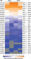
Fig. 4.. Secretome profiling of A. bisporus lignin treatments.
Protein IDs and log2-transformed IBAQ intensities of enzymes active on lignin and aromatics in secretomes of biological duplicates (coded A15I and A15II) in analytical duplicates of A. bisporus cultivated on nonlabeled lignin in minimal medium (secretomes of 12CLg + Ab; data S1). CAZyme family and subfamily, protein ID, and putative functions are provided if available [according to Joint Genome Institute identifier “jgi|Agabi_varbisH97_2” and Morin et al. (11)]. MCO, multicopper oxidase; UPO, unspecific peroxygenase; ODC, oxalate decarboxylase; MnP, manganese peroxidase; GLOX, glyoxal oxidase; AAO, aryl alcohol oxidase; GDH, glucose dehydrogenase; GST, glutathione S-transferase.
Fig. 5.
Fig. 5.. 13C-fractional labeling of FBM compounds.
Fatty acids and ergosterol (A), amino acids (B), carbohydrates (C), of A. bisporus (Ab) FBM from 12C*Lg + Ab (orange bars; 12C-lignin “spiked” used as carbon source), and from 13CLg + Ab (turquoise bars; 13C-lignin used as carbon source). All results are the average of biological duplicates, and error bars represent the SDs. The graphs on the right side indicate maximal labeling possible, calculated from FBM increase and mycelium seed used, for 13CLg + Ab (turquoise), and labeled spike used for 12C*Lg + Ab (orange).
Fig. 6.
Fig. 6.. Extracellular lignin active enzymes and intracellular pathways and conversion of assimilated lipids, arabinose/xylose, glucose, and lignin degradation products in a fungal cell.
Metabolic pathways show key intermediates where they branch off for biosynthesis of amino acids, hexoses/riboses, and lipids. Amino acids are shown in their one-letter codes (magenta). Key-branching points for biosynthesis of riboses, β-glucan, and chitin are glucose-6-phosphate (G6P) and fructose-6-phosphate (F6P; green). These fungal components/fibers can be hydrolyzed and result in hexoses and riboses (green) for determining the 13C-enrichment. Lipids and steroids are marked in yellow and 13C-fractional enrichment was tested in fatty acids and ergosterol. Extracellularly, lignin is depolymerized by lignin active enzymes (dark blue), which have been investigated in A. bisporus secretome cultivated on nonlabeled lignin. MCO, multicopper oxidase; PPP, pentose phosphate pathway; EMP, Embden-Meyerhof-Parnas pathway/glycolysis; GNG, gluconeogenesis; TCA cycle, tricarboxylic acid cycle; G3P, glycerol-3-phosphate; 3PG, 3-phosphoglycerate; PEP, phosphoenolpyruvate; X5P, xylose-5-phosphate; R5P, ribose-5-phosphate; αKG, α-ketoglutarate; Oa, oxaloacetate; PW, pathway. The figure was created with BioRender (BioRender.com). Metabolic pathways were based on the Kyoto Encyclopedia of Genes and Genomes (KEGG) database (33, 34), and proposed end products of aromatic-ring cleavage pathways are based on the schemes proposed by Holesova et al. (31), Lubbers et al. (29), del Cerro et al. (10), Kijpornyongpan et al. (5), and Patyshakuliyeva et al. (47).

References

    1. Liang C., Schimel J. P., Jastrow J. D., The importance of anabolism in microbial control over soil carbon storage. Nat. Microbiol. 2, 17105 (2017). - PubMed
    1. Tao F., Huang Y., Hungate B. A., Manzoni S., Frey S. D., Schmidt M. W. I., Reichstein M., Carvalhais N., Ciais P., Jiang L., Lehmann J., Wang Y. P., Houlton B. Z., Ahrens B., Mishra U., Hugelius G., Hocking T. D., Lu X., Shi Z., Viatkin K., Vargas R., Yigini Y., Omuto C., Malik A. A., Peralta G., Cuevas-Corona R., Di Paolo L. E., Luotto I., Liao C., Liang Y. S., Saynes V. S., Huang X., Luo Y., Microbial carbon use efficiency promotes global soil carbon storage. Nature 618, 981–985 (2023). - PMC - PubMed
    1. Kang X., Kirui A., Dickwella Widanage M. C., Mentink-Vigier F., Cosgrove D. J., Wang T., Lignin-polysaccharide interactions in plant secondary cell walls revealed by solid-state NMR. Nat. Commun. 10, 347 (2019). - PMC - PubMed
    1. Cragg S. M., Beckham G. T., Bruce N. C., Bugg T. D. H., Distel D. L., Dupree P., Etxabe A. G., Goodell B. S., Jellison J., McGeehan J. E., McQueen-Mason S. J., Schnorr K., Walton P. H., Watts J. E. M., Zimmer M., Lignocellulose degradation mechanisms across the tree of life. Curr. Opin. Chem. Biol. 29, 108–119 (2015). - PMC - PubMed
    1. Kijpornyongpan T., Schwartz A., Yaguchi A., Salvachúa D., Systems biology-guided understanding of white-rot fungi for biotechnological applications: A review. iScience 25, 104640 (2022). - PMC - PubMed

Publication types

Supplementary concepts