. 2024 Jun:226:105888.
doi: 10.1016/j.antiviral.2024.105888. Epub 2024 Apr 18.

An allosteric inhibitor of sirtuin 2 blocks hepatitis B virus covalently closed circular DNA establishment and its transcriptional activity

Affiliations

An allosteric inhibitor of sirtuin 2 blocks hepatitis B virus covalently closed circular DNA establishment and its transcriptional activity

Liudi Tang et al. Antiviral Res. 2024 Jun.

Abstract

296 million people worldwide are predisposed to developing severe end-stage liver diseases due to chronic hepatitis B virus (HBV) infection. HBV forms covalently closed circular DNA (cccDNA) molecules that persist as episomal DNA in the nucleus of infected hepatocytes and drive viral replication. Occasionally, the HBV genome becomes integrated into host chromosomal DNA, a process that is believed to significantly contribute to circulating HBsAg levels and HCC development. Neither cccDNA accumulation nor expression from integrated HBV DNA are directly targeted by current antiviral treatments. In this study, we investigated the antiviral properties of a newly described allosteric modulator, FLS-359, that targets sirtuin 2 (SIRT2), an NAD+-dependent deacylase. Our results demonstrate that SIRT2 modulation by FLS-359 and by other tool compounds inhibits cccDNA synthesis following de novo infection of primary human hepatocytes and HepG2 (C3A)-NTCP cells, and FLS-359 substantially reduces cccDNA recycling in HepAD38 cells. While pre-existing cccDNA is not eradicated by short-term treatment with FLS-359, its transcriptional activity is substantially impaired, likely through inhibition of viral promoter activities. Consistent with the inhibition of viral transcription, HBsAg production by HepG2.2.15 cells, which contain integrated HBV genomes, is also suppressed by FLS-359. Our study provides further insights on SIRT2 regulation of HBV infection and supports the development of potent SIRT2 inhibitors as HBV antivirals.

Keywords: Antiviral; Hepatitis B virus; SIRT2; Viral transcription; cccDNA synthesis.


Conflict of interest statement

Declaration of competing interest The authors declare the following financial interests/personal relationships which may be considered as potential competing interests.

Figures

Fig. 1.
Fig. 1.
FLS-359 blocks HBV infection in primary human hepatocytes. (A) Schematic representation of the virus infection and compound treatment schedule. PXB-cells were mock-infected or infected with HBV (500 GE s/cell), treated with DMSO or FLS-359 in DMSO from 4 h prior to the infection to 6 dpi, or with MyrB 4 h prior to the infection to 1 dpi. Cells and culture supernatant were harvested at 6 dpi. (B) HBV cccDNA was quantified by qPCR and normalized to the infected- and DMSO-treated group. Mean ± SD is shown (n = 3, each with 2 qPCR technical repeats). (C) Total HBV RNA transcripts were quantified by RT-qPCR and normalized to cellular β-actin RNA. Data are presented as HBV RNA levels relative to the infected- and DMSO-treated group. Mean ± SD is shown (n = 3). HBsAg (D) and HBeAg (E) secreted into culture supernatant from 4 to 6 dpi were quantified by CLIA assay. Mean ± SD is shown (n = 4). (F) PXB cells were treated with DMSO or FLS-359 in DMSO at indicated concentrations for 8 days; cells were washed with PBS and fixed with 2% paraformaldehyde, followed by DAPI staining. Cell viability was determined by DAPI cell count, and normalized to the DMSO-treated group. Mean ± SD is shown (n = 2). *p < 0.05; **p < 0.01; ***p < 0.001.
Fig. 2.
Fig. 2.
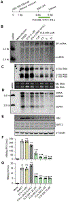
FLS-359 reduces HBV cccDNA establishment in C3A-NTCP cells. (A) Schematic representation of the virus infection and compound treatment schedule. C3A-NTCP cells were mock-infected or infected with HBV (2000 GE s/cell), and treated with DMSO or with drugs in DMSO at indicated concentrations and time frames. Cells and culture supernatant were harvested at 6 dpi. (B) Hirt DNA was extracted and analyzed by Southern blot assay using DIG-labeled full length HBV DNA probe. The cccDNA and DP-rcDNA are labeled. (C) Total RNA was resolved by Northern blot assay using DIG-labeled full length HBV DNA probe. HBV 3.5 kb RNA, 2.4 kb RNA, and 2.1 kb RNA are labeled. 18 S and 28 S ribosomal RNA serve as internal controls. (D) Replicative cytoplasmic core DNA was resolved by Southern blot assay using DIG-labeled full length HBV DNA probe. rcDNA, dslDNA, and ssDNA are labeled. (E) The levels of HBV core protein (HBc), SIRT2, and α-tubulin were determined by Western blot assay. (F-G) HBeAg and HBsAg secreted into culture medium from 4 to 6 dpi were quantified by CLIA assay. Mean ± SD is shown (n = 4). *p < 0.05, **p < 0.01, ***p < 0.001.
Fig. 3.
Fig. 3.
FLS-359 reduces HBV RNA production in HBV-infected primary human hepatocytes. (A) Schematic representation of the virus infection and compound treatment schedule. PXB cells were mock-infected or infected with HBV (500 GE s/cell). From 4 dpi, 1 μM ETV was maintained in cultures to block the potential replenishment of cccDNA through the recycling pathway, and cells were treated with DMSO or FLS-359 in DMSO at indicated concentrations until harvesting cells and culture supernatants at 10 dpi. (B) Hirt DNA was extracted, and HBV cccDNA was quantified by qPCR and normalized to the DMSO-treated group. Mean ± SD is shown (n = 3, each with 2 qPCR technical repeats). (C) Total RNA was extracted from cells, and HBV RNA was quantified by RT-qPCR and normalized to cellular β-actin RNA amounts. Data are presented as HBV RNA levels relative to the DMSO-treated group. Mean ± SD is shown (n = 3). HBsAg (D) and HBeAg (E) secreted into culture medium from 7 to 10 dpi were quantified by CLIA assay. Mean ± SD is shown (n = 4). ***p < 0.001.
Fig. 4.
Fig. 4.
FLS-359 does not induce degradation of existing cccDNA, but reduces HBV RNA accumulation in C3A-NTCP cells. (A) Schematic representation of the virus infection and compound treatment schedule. C3A-NTCP cells were mock-infected or infected with HBV (2000 GE s/cell). From 4 to 6 dpi, cells were treated with DMSO or drugs in DMSO at indicated concentrations. Cells and culture supernatant were harvested at 6 dpi. (B) Hirt DNA was extracted and resolved by Southern blot assay using DIG-labeled full length HBV DNA probe. cccDNA and DP-rcDNA are labeled. (C) Total RNA was extracted from cells and analyzed by Northern blot assay using DIG-labeled full length HBV DNA probe. HBV 3.5 kb RNA, 2.4 kb RNA, and 2.1 kb RNA are labeled. 18 S and 28 S ribosomal RNA serve as internal controls. (D) Replicative cytoplasmic coreDNA was extracted and analyzed by Southern blot assay using DIG-labeled full length HBV DNA probe. rcDNA, dslDNA, and ssDNA were denoted. (E) The levels of HBV core protein (HBc), SIRT2, and α-tubulin were determined by Western blot assay. (F-G) Secreted HBeAg and HBsAg that accumulated from 4 to 6 dpi were quantified by CLIA assay. Mean ± SD is shown (n = 4). **p < 0.01, ***p < 0.001.
Fig. 5.
Fig. 5.
FLS-359 hampers HBV RNA, DNA and protein production in HepG2.2.15 cells. (A) Schematic representation of the compound treatment. HepG2.2.15 cells were treated with DMSO, ETV (1 μM) or FLS-359 at indicated concentrations. Cells and culture supernatant were harvested 2 days after drug treatment. (B) Total RNA was extracted from cells and analyzed by Northern blot assay using a32P-labeled full-length minus strand HBV riboprobe. HBV 3.5 kb RNA and 2.4/2.1 kb RNA species are labeled. 18 S and 28 S ribosomal RNA serve as loading controls. (C) Replicative cytoplasmic core DNA was analyzed by Southern blot assay using a32P-labeled full-length plus strand HBV riboprobe. rcDNA and ssDNA are labeled. (D) HBV capsid was detected by using a particle gel assay. β-actin protein levels were determined by Western blot assay using same number of cells as used to quantify capsids. (E-F) Secreted HBeAg and HBsAg that accumulated during the 2-day treatment was quantified by CLIA assay. Mean ± SD is shown (n = 4). **p < 0.01, ***p < 0.001.
Fig. 6.
Fig. 6.
FLS-359 leads to rapid reduction of viral RNA and antigen in HBV- infected hepatocytes and in HepG2.2.15 cells. (A–B) Schematic representation of the virus infection and compound treatment schedule. C3A-NTCP cells were infected with HBV (500 GE s/cell). At 5 dpi, cells were treated with DMSO or 10 μM FLS-359 in DMSO. Culture supernatants were collected after the indicated time periods of drug treatment, and HBeAg levels were quantified by CLIA assay. (C-D) Schematic representation of the virus infection and compound treatment schedule. C3A-NTCP cells were infected with HBV (500 GE s/cell). Cells were harvested at 7 dpi following the indicated periods of treatment with 10 μM FLS-359, and total cell RNA was prepared. Total HBV RNA levels were quantified by qRT-PCR, normalized to β-actin RNA, and results are presented as fold-change relative to the untreated group. (E-F) Schematic representation of the compound treatment schedule. HepG2.2.15 cells were treated with DMSO or 10 μM FLS-359 in DMSO. Culture supernatants were collected after indicated periods of drug treatment, and HBsAg levels were quantified by CLIA assay. (G-H) Schematic representation of the compound treatment schedule. HepG2.2.15 cells were harvested after the indicated periods of treatment with 10 μM FLS-359 treatment, and total cell RNA was prepared. Total HBV RNA levels were quantified by qRT-PCR, normalized to β-actin RNA, and results are presented as fold-change relative to the untreated group. For all panels, mean ± SD is shown (n = 4). *p < 0.05, **p < 0.01, ***p < 0.001.
Fig. 7.
Fig. 7.
FLS-359 suppresses HBV RNA production. (A) HepG2 cells infected with Ad-HBV2.1 and cultured in the presence or absence of Dox were harvested at 3 dpi and total cellular RNA was extracted. HBV 2.1 kb RNA was quantified by qRT-PCR, normalized to β-actin RNA, and results are presented as fold-change relative to the Dox-group. Mean ± SD is shown (n = 3). (B) Schematic representation of the virus infection and compound treatment schedule. HepG2 cells were infected with Ad-HBV2.1, and cultured in the absence of doxycycline (Dox) for 3 days before shutting down the viral RNA production by switching to medium with Dox (2 μg/ml). Cells were harvested at various times after treatment with DMSO, FLS-359 (10 μM in DMSO) or RG-7834 (10 μM in DMSO). (C) HBV 2.1 kb RNA was measured by qRT-PCR, normalized to β-actin RNA. Mean ± SD is shown (n = 3). (D) HepG2 cells were transfected with reporters containing each of the four HBV promoters or a control HCMV promoter (IEp) for 24 h and then treated with FLS-359 at indicated concentrations for another 24 h before performing luciferase assays. The absolute luciferase levels for drug-treated cells are presented as a percentage of the level for cells treated with DMSO. Mean ± SD is shown (n = 3). *p < 0.05, **p < 0.01, ***p < 0.001.

Similar articles

Cited by

References

    1. Allweiss L, Giersch K, Pirosu A, Volz T, Muench RC, Beran RK, Urban S, Javanbakht H, Fletcher SP, Lutgehetmann M, Dandri M, 2022. Therapeutic shutdown of HBV transcripts promotes reappearance of the SMC5/6 complex and silencing of the viral genome in vivo. Gut 71, 372–381. 10.1136/gutjnl-2020-322571. - DOI - PMC - PubMed
    1. Alter H, Block T, Brown N, Brownstein A, Brosgart C, Chang KM, Chen PJ, Chisari FV, Cohen C, El-Serag H, Feld J, Gish R, Glenn J, Greten T, Guo H, Guo JT, Hoshida Y, Hu J, Kowdley KV, Li W, Liang J, Locarnini S, Lok AS, Mason W, McMahon B, Mehta A, Perrillo R, Revill P, Rice CM, Rinaudo J, Schinazi R, Seeger C, Shetty K, Tavis J, Zoulim F, 2018. A research agenda for curing chronic hepatitis B virus infection. Hepatology 67, 1127–1131. 10.1002/hep.29509. - DOI - PMC - PubMed
    1. Bill CA, Summers J, 2004. Genomic DNA double-strand breaks are targets for hepadnaviral DNA integration. Proc. Natl. Acad. Sci. U. S. A 101, 11135–11140. 10.1073/pnas.0403925101. - DOI - PMC - PubMed
    1. Bloom K, Maepa MB, Ely A, Arbuthnot P, 2018. Gene therapy for chronic HBV-can we eliminate cccDNA? Genes (Basel) 9. 10.3390/genes9040207. - DOI - PMC - PubMed
    1. Boyd A, Lacombe K, Lavocat F, Maylin S, Miailhes P, Lascoux-Combe C, Delaugerre C, Girard PM, Zoulim F, 2016. Decay of ccc-DNA marks persistence of intrahepatic viral DNA synthesis under tenofovir in HIV-HBV co-infected patients. J. Hepatol 65, 683–691. 10.1016/j.jhep.2016.05.014. - DOI - PubMed

MeSH terms

Substances

LinkOut - more resources