

Ageing of Plasmodium falciparum malaria sporozoites alters their motility, infectivity and reduces immune activation in vitro
- PMID: 38641838
- PMCID: PMC11027264
- DOI: 10.1186/s12936-024-04946-7
Ageing of Plasmodium falciparum malaria sporozoites alters their motility, infectivity and reduces immune activation in vitro
Abstract
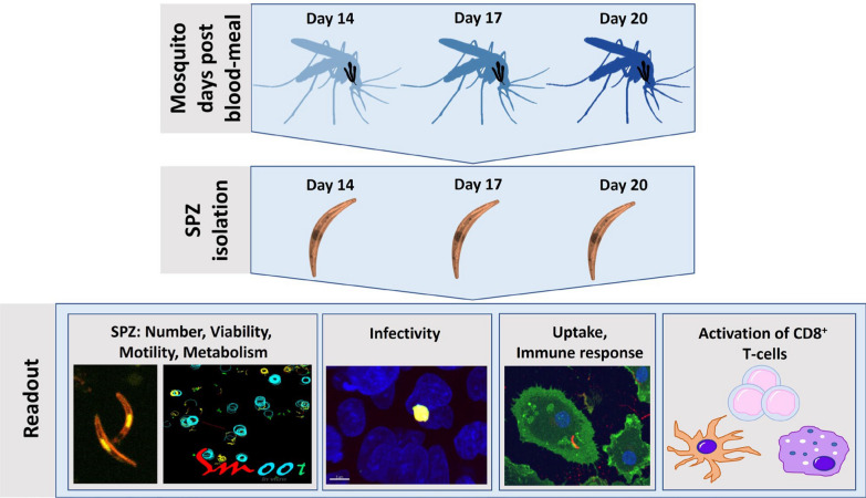
Background: Sporozoites (SPZ), the infective form of Plasmodium falciparum malaria, can be inoculated into the human host skin by Anopheline mosquitoes. These SPZ migrate at approximately 1 µm/s to find a blood vessel and travel to the liver where they infect hepatocytes and multiply. In the skin they are still low in number (50-100 SPZ) and vulnerable to immune attack by antibodies and skin macrophages. This is why whole SPZ and SPZ proteins are used as the basis for most malaria vaccines currently deployed and undergoing late clinical testing. Mosquitoes typically inoculate SPZ into a human host between 14 and 25 days after their previous infective blood meal. However, it is unknown whether residing time within the mosquito affects SPZ condition, infectivity or immunogenicity. This study aimed to unravel how the age of P. falciparum SPZ in salivary glands (14, 17, or 20 days post blood meal) affects their infectivity and the ensuing immune responses.
Methods: SPZ numbers, viability by live/dead staining, motility using dedicated sporozoite motility orienting and organizing tool software (SMOOT), and infectivity of HC-04.j7 liver cells at 14, 17 and 20 days after mosquito feeding have been investigated. In vitro co-culture assays with SPZ stimulated monocyte-derived macrophages (MoMɸ) and CD8+ T-cells, analysed by flow cytometry, were used to investigate immune responses.
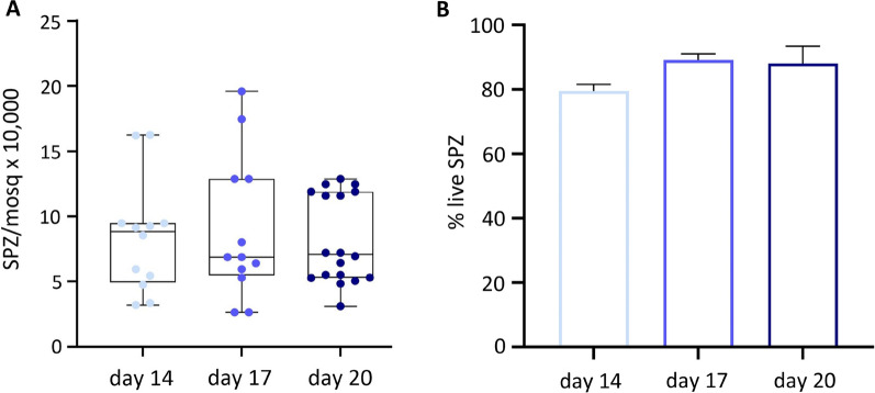
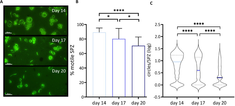
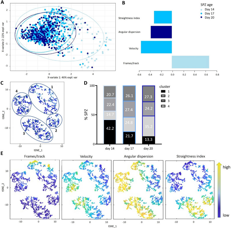
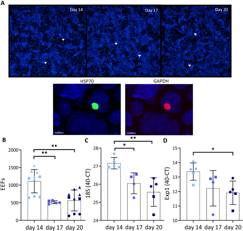
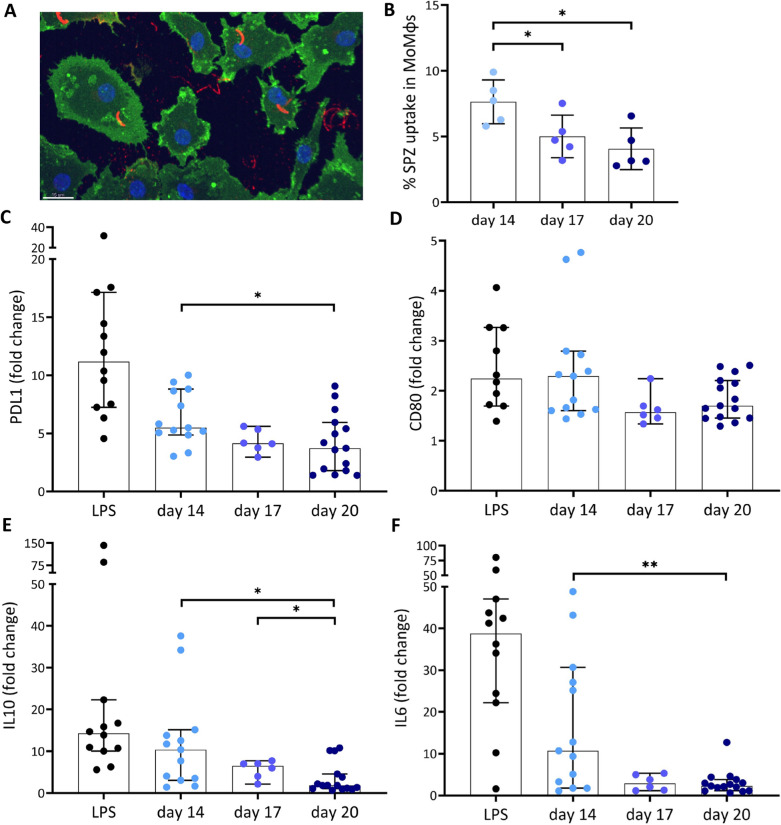
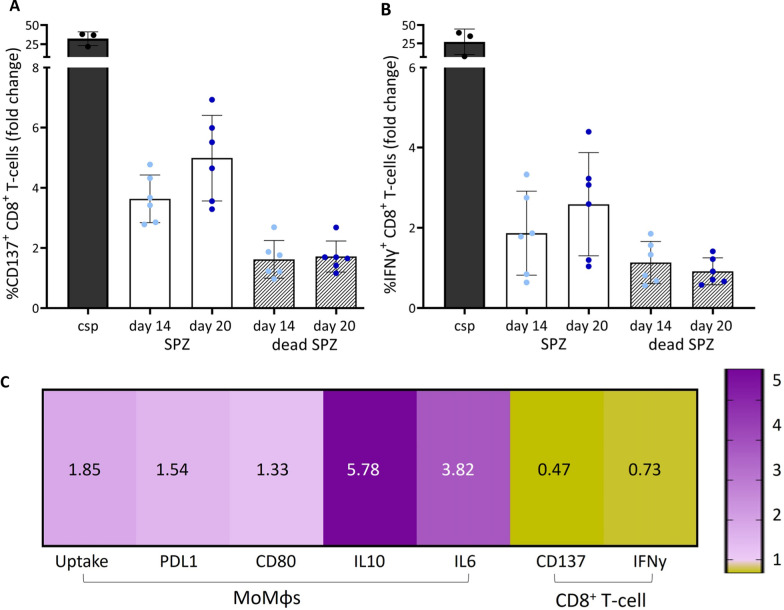
Results: SPZ age did not result in different SPZ numbers or viability. However, a markedly different motility pattern, whereby motility decreased from 89% at day 14 to 80% at day 17 and 71% at day 20 was observed (p ≤ 0.0001). Similarly, infectivity of day 20 SPZ dropped to ~ 50% compared with day 14 SPZ (p = 0.004). MoMɸ were better able to take up day 14 SPZ than day 20 SPZ (from 7.6% to 4.1%, p = 0.03) and displayed an increased expression of pro-inflammatory CD80, IL-6 (p = 0.005), regulatory markers PDL1 (p = 0.02), IL-10 (p = 0.009) and cytokines upon phagocytosis of younger SPZ. Interestingly, co-culture of these cells with CD8+ T-cells revealed a decreased expression of activation marker CD137 and cytokine IFNγ compared to their day 20 counterparts. These findings suggest that older (day 17-20) P. falciparum SPZ are less infectious and have decreased immune regulatory potential.
Conclusion: Overall, this data is a first step in enhancing the understanding of how mosquito residing time affects P. falciparum SPZ and could impact the understanding of the P. falciparum infectious reservoir and the potency of whole SPZ vaccines.
Keywords: Plasmodium falciparum; Immunogenicity; Malaria; Motility; Sporozoites.
© 2024. The Author(s).
Conflict of interest statement
The author(s) declare that they have no competing interest.
Figures
Similar articles
-
Clustering and Erratic Movement Patterns of Syringe-Injected versus Mosquito-Inoculated Malaria Sporozoites Underlie Decreased Infectivity.mSphere. 2021 Apr 7;6(2):e00218-21. doi: 10.1128/mSphere.00218-21. mSphere. 2021. PMID: 33827910 Free PMC article.
-
Detection of CD4+CD45RO+ T lymphocytes producing IL-4 in response to antigens on Plasmodium falciparum erythrocytes: an in vitro correlate of protective immunity induced with attenuated Plasmodium falciparum sporozoites.Cell Immunol. 1997 Sep 15;180(2):143-52. doi: 10.1006/cimm.1997.1186. Cell Immunol. 1997. PMID: 9341744
-
Sporozoite motility as a quantitative readout for anti-CSP antibody inhibition.Sci Rep. 2022 Oct 13;12(1):17194. doi: 10.1038/s41598-022-22154-8. Sci Rep. 2022. PMID: 36229488 Free PMC article.
-
Current Challenges in the Identification of Pre-Erythrocytic Malaria Vaccine Candidate Antigens.Front Immunol. 2020 Feb 21;11:190. doi: 10.3389/fimmu.2020.00190. eCollection 2020. Front Immunol. 2020. PMID: 32153565 Free PMC article. Review.
-
The whole parasite, pre-erythrocytic stage approach to malaria vaccine development: a review.Curr Opin Infect Dis. 2013 Oct;26(5):420-8. doi: 10.1097/QCO.0000000000000002. Curr Opin Infect Dis. 2013. PMID: 23982233 Review.
References
-
- WHO. World Malaria Report 2022. Geneva: World Health Organization; 2022
-
- Walther M. Advances in vaccine development against the pre-erythrocytic stage of Plasmodium falciparum malaria. Expert Rev Vaccines. 2006;5:81–93. - PubMed
MeSH terms
Substances
Grants and funding
LinkOut - more resources
Full Text Sources
Medical
Research Materials
