. 2024 Aug 28;75(16):4802-4821.
doi: 10.1093/jxb/erae180.

OsRH52A, a DEAD-box protein, regulates functional megaspore specification and is required for embryo sac development in rice

Affiliations

OsRH52A, a DEAD-box protein, regulates functional megaspore specification and is required for embryo sac development in rice

Jinghua Huang et al. J Exp Bot. .

Abstract

The development of the embryo sac is an important factor that affects seed setting in rice. Numerous genes associated with embryo sac (ES) development have been identified in plants; however, the function of the DEAD-box RNA helicase family genes is poorly known in rice. Here, we characterized a rice DEAD-box protein, RH52A, which is localized in the nucleus and cytoplasm and highly expressed in the floral organs. The knockout mutant rh52a displayed partial ES sterility, including degeneration of the ES (21%) and the presence of a double-female-gametophyte (DFG) structure (11.8%). The DFG developed from two functional megaspores near the chalazal end in one ovule, and 3.4% of DFGs were able to fertilize via the sac near the micropylar pole in rh52a. RH52A was found to interact with MFS1 and ZIP4, both of which play a role in homologous recombination in rice meiosis. RNA-sequencing identified 234 down-regulated differentially expressed genes associated with reproductive development, including two, MSP1 and HSA1b, required for female germline cell specification. Taken together, our study demonstrates that RH52A is essential for the development of the rice embryo sac and provides cytological details regarding the formation of DFGs.

Keywords: Oryza sativa; Cell specification; DEAD-box RNA helicase; chalazal functional megaspore; double-female-gametophyte; embryo sac; rice.


Conflict of interest statement

The authors declare that they have no conflicts of interest in relation to this work.

Figures

Fig. 1.
Fig. 1.
Homology analysis and subcellular localization of OsRH52A. (A) Multiple amino acid sequence alignment of proteins from the DEAD-box RNA helicase family in Oryza sativa (OsRH52A, OsRH52C), Arabidopsis thaliana (AtRH11), Triticum aestivum (TaRH11), Zea mays (ZmRH11), Setaria italic (SiRH11), and Sorghum bicolor (SbRH52A). The red lines indicate the DEXDc and HELICc domains of OsRH52A, whilst the eight motifs of DEAD-box RNA helicase are indicated by the black rectangles. (B) The subcellular localization of the OsRH52A protein in rice protoplasts. GFP, green fluorescence protein. ‘Merge’ is the overlapped image of GFP, the nuclear marker NLS-mCherry, and the bright field. Scale bars are 20 μm. (C) The subcellular localization of the OsRH52A protein in leaves of Nicotiana benthamiana. NLS-mKATE was used as the nuclear marker. Scale bars are 40 μm.
Fig. 2.
Fig. 2.
Expression patterns of RH52A in different tissues of wild type Nipponbare rice. (A) Relative expression of RH52A in the roots, stem, leaves, and developing spikelets. PM, anther and ovary at the pre-meiosis stage; MA, anther at meiosis stage; SCP1, early microspore stage; SCP2, middle microspore stage; SCP3, late microspore stage; BCP1, early bicellular pollen stage; BCP2, late bicellular pollen stage; M, mature anther. Ov1, ovary at meiosis of embryo sac (ES); Ov2, ovary at functional megaspore stage of ES; Ov3, ovary at mitosis of ES; Ov4, ovary of mature ES. (B) Histochemical staining of developing spikelets (top) and their corresponding reproductive organs (bottom) expressing the GUS reporter gene driven by the RH52A promoter. The length of the developing spikelets is indicated below the images. Scale bars are 2 mm.
Fig. 3.
Fig. 3.
CRISPR/Cas9 mutation of rice RH52A and analysis of its effects on seed setting and pollen fertility. (A) Nucleotide alignments of the CRISPR/Cas9 target sites in the Nipponbare wild type (WT) and the two mutants rh52a-m1 and rh52a-m2. In the schematic diagram, the lines represent the introns and the black boxes show the exons; grey boxes indicate the untranslated regions. The two target sites are indicated. (B) Amino acid alignments of RH52A in the WT and the rh52a-m1 and rh52a-m2 mutants. (C) Seed setting rate of the WT and the rh52a-m1 and rh52a-m2 mutants. Values are means (±SD), n=15. Significant differences compared with the WT were determined using Student’s t-test: **P<0.01. (D) Frequency distribution of F2 populations from the cross between rh52a-m2 and the WT. (E) Seed setting rates of 32 randomly selected lines from the F2 population that were genotyped by Sanger sequencing. (F) Pollen fertility of the WT and three randomly selected homozygous lines (yy) from the F2 population. Values are means (±SD), n=3. (G) Pollen fertility of the WT and the three homozygous lines (yy) using I2-KI staining assays. Scale bars are 100 μm.
Fig. 4.
Fig. 4.
Representative images of various abnormal embryo sacs of the rice wild type (WT) and the rh52a-m2 mutant. Red embryo sacs were obtained by whole-mount eosin B-staining/confocal laser-scanning microscopy (WE-CLSM). Blue embryo sacs were obtained from semi-thin sections stained with Toluidine blue O. Wa and Wb show normal mature embryo sacs of the WT. Ma1 and Mb1 show embryo sac degeneration of the mutant. EA, egg apparatus (consisting of an egg cell and two synergids); PN, polar nuclei; AC, antipodal cells. Ma2 and Mb2 show stacked embryo sacs of the mutants. Ma3 and Mb3 show embryo sacs of the mutants without female germ units. Ma4 and Mb4 show embryo sacs of the mutants without egg apparatus. Wc shows the megasporocyte of the WT. Mc1 to Mc4 show multiple megasporocytes of the mutants. The arrows in Wc and Mc1–4 indicate the nuclei. All scale bars are 40 μm.
Fig. 5.
Fig. 5.
Embryo sac developmental process from the functional megaspore (FM) stage to the tetra-nucleate in the rice wild type (WT) and the rh52a-m2 mutant. Red embryo sacs were obtained by whole-mount eosin B-staining/confocal laser-scanning microscopy (WE-CLSM). Blue embryo sacs were obtained from semi-thin sections stained with Toluidine blue O. Wa shows the FM of the WT. Ma1 to Ma3 show the double FMs of the mutants. Wb shows the mono-nucleate embryo sac of the WT. Mb1–Mb3 show the double mono-nucleate embryo sacs (MN) of the mutants. Wc shows the bi-nucleate embryo sacs of the WT. Mc1–Mc3, and Md3 show the double bi-nucleate embryo sacs (BN) of the mutants. Wd shows the tetra-nucleate embryo sacs of the WT. Md1 and Md2 show the double tetra-nucleate embryo sacs (TN) of the mutants. All scale bars are 40 μm.
Fig. 6.
Fig. 6.
Embryo sac developmental process from the eight-nucleate stage to 3 days after fertilization (DAF) in the rice wild type (WT) and the rh52a-m2 mutants. Red embryo sacs were obtained by whole-mount eosin B-staining/confocal laser-scanning microscopy (WE-CLSM). Blue embryo sacs were obtained from semi-thin sections stained with Toluidine blue O staining. Wa shows the eight-nucleate embryo sac of the WT. Ma1–Ma3 show the double eight-nucleate embryo sacs (EN) of the mutants. Wb shows the mature embryo sac of the WT. EA, egg apparatus (consisting of an egg cell and two synergids); PN, polar nuclei; AC, antipodal cells. Mb1–Mb3 show the double embryo sacs (SAC) of the mutants. Wc shows the embryo sac of the WT at 1 DAF. Mc1–Mc3 show double embryo sacs of the mutants at 1 DAF. Wd shows the embryo sac of the WT at 3 DAF. Md1 shows the double embryo sacs of the mutant at 3 DAF. Wd and Md1 are compound images constructed from individual segments. Scale bars: Wa, Ma1–Ma3, Wb, Mb1–Mb3=40 μm; Wc, Mc1–Mc3=50 μm; Wd, Md1=100 μm.
Fig. 7.
Fig. 7.
OsRH52A physically interacts with OsMFS1 and OsZIP4. (A) Yeast two-hybrid (Y2H) assays showing no toxicity and autoactivation of the OsRH52A protein. (B) Y2H assays showing the interaction of OsRH52A with OsMFS1 and with OsZIP4. (C) Bimolecular fluorescence complementation assays of OsRH52A with OsMFS1 and OsZIP4 in leaf epidermal cells of Nicotiana benthamiana. Scale bars are 40 μm. (D) Luciferase complementation imaging assays showing the interaction of OsRH52A with OsMFS1 and with OsZIP4 in N. benthamiana leaves.
Fig. 8.
Fig. 8.
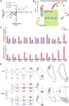
RNA-sequencing analysis and a proposed scheme for the development of double-female-gametophytes in the rice wild type and rh52a mutant. (A) Correlation between qRT-PCR analysis and the RNA-seq results for 19 selected genes. The qRT-PCR analysis was carried out using either UBI as the reference gene (red circles) or CPI (green circles). (B) Mapping of 17 down-regulated differentially expressed genes to ubiquitin degradation pathways in the rh52a-m2 mutant using the MapMan tool. The relative expression of each of the 17 genes is indicated according to the heatmap scale. (C) The relative expression of genes related to megagametophytes in the wild type (WT) and the mutant at the functional megaspore stage, as determined by qRT-PCR Using either UBI or CPI as the reference gene. (D) Schematic diagram of the double-female-gametophyte (DFG) developmental process from the functional megaspore to the mature stage, and at 1 day after fertilization (DAF) and 3 DAF. FM, functional megaspore; MN, mono-nucleate embryo sac (ES); BN, bi-nucleate ES; TN, tetra-nucleate ES; EN, eight-nucleate ES. AC, antipodal cells. The embryo sac at the mitosis stage in the wild type (WT) is indicated by the dash box. For the rh52a mutant, the dashed red box indicates the process of synchronous division in each sac from DFG at the mitosis stage, whilst the blue box indicates the process of asynchronous division.

Similar articles

Cited by

References

    1. Ao C-Q. 2013. Developmental origins of the conjoined twin mature embryo sacs in Smilax davidiana, with special notes on the formation of their embryos and endosperms. American Journal of Botany 100, 2509–2515. - PubMed
    1. Awasthi A, Paul P, Kumar S, Verma SK, Prasad R, Dhaliwal HS.. 2012. Abnormal endosperm development causes female sterility in rice insertional mutant OsAPC6. Plant Science 183, 167–174. - PubMed
    1. Cao GQ, Gu HH, Jiang WJ, et al.. 2022. BrDHC1, a novel putative DEAD-box helicase gene, confers drought tolerance in transgenic Brassica rapa. Horticulturae 8, 707.
    1. Capel C, Albaladejo I, Egea I, et al.. 2020. The res (restored cell structure by salinity) tomato mutant reveals the role of the DEAD-box RNA helicase SlDEAD39 in plant development and salt response. Plant, Cell & Environment 43, 1722–1739. - PMC - PubMed
    1. Chang YX, Gong L, Yuan WY, Li XW, Chen GX, Li XH, Zhang QF, Wu CY.. 2009. Replication protein A (RPA1a) is required for meiotic and somatic DNA repair but is dispensable for DNA replication and homologous recombination in rice. Plant Physiology 151, 2162–2173. - PMC - PubMed

LinkOut - more resources