. 2024 Apr;41(2):163-174.
doi: 10.1007/s10719-024-10150-1. Epub 2024 Apr 20.

Marine sulfated glycans inhibit the interaction of heparin with S-protein of SARS-CoV-2 Omicron XBB variant

Affiliations

Marine sulfated glycans inhibit the interaction of heparin with S-protein of SARS-CoV-2 Omicron XBB variant

Peng He et al. Glycoconj J. 2024 Apr.

Abstract

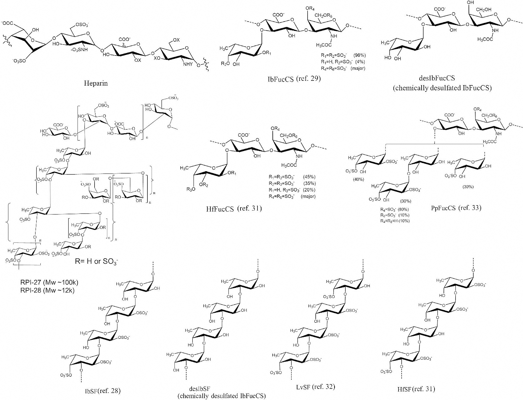
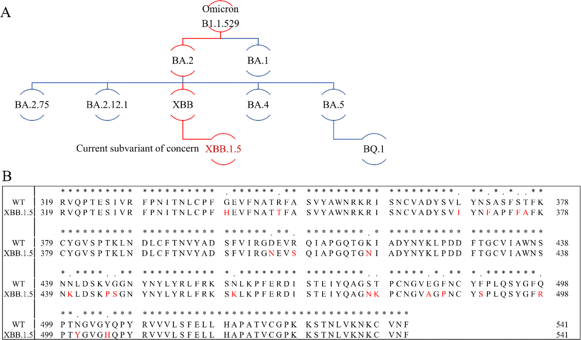
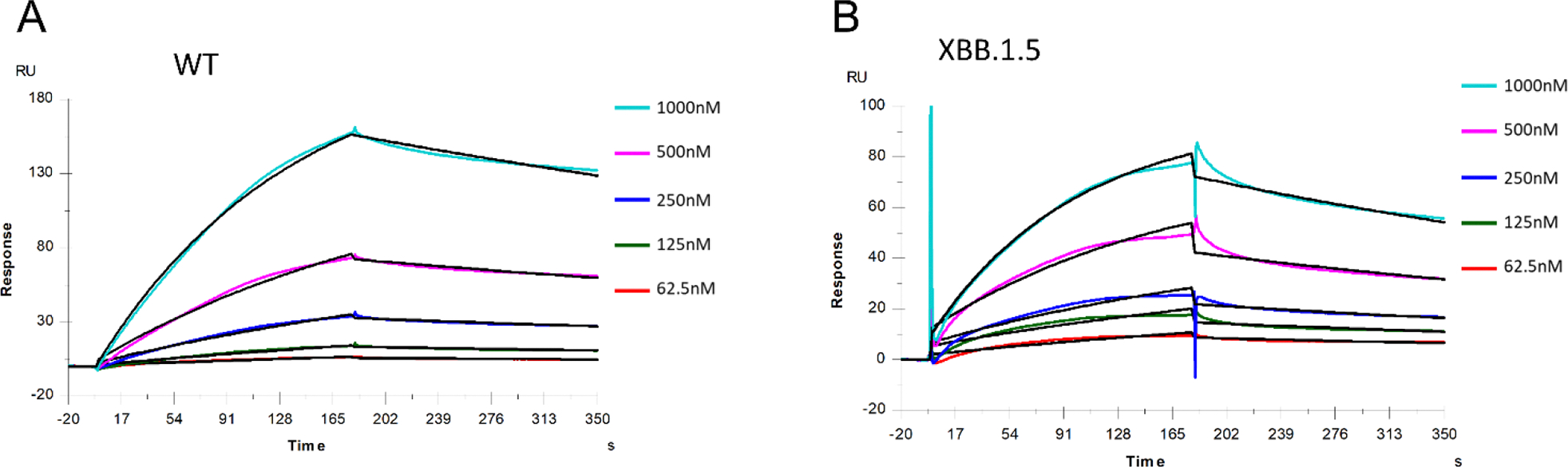
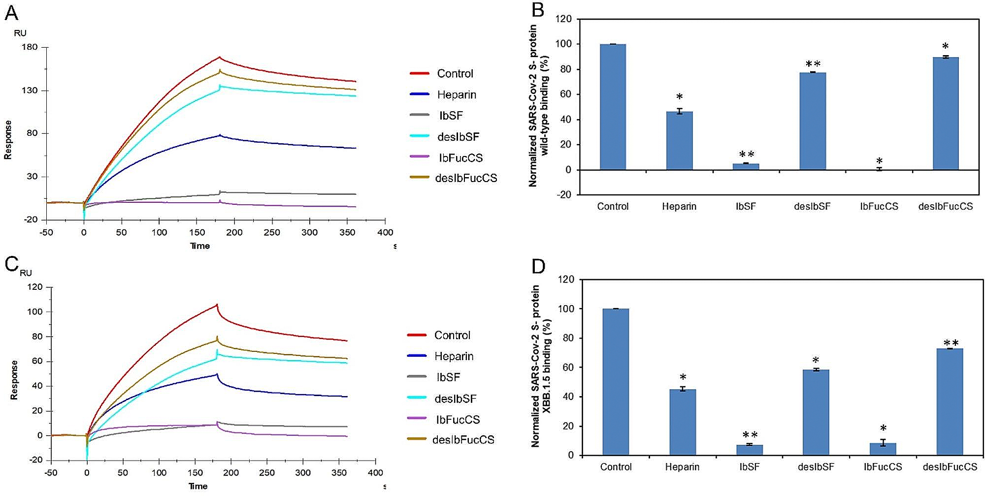
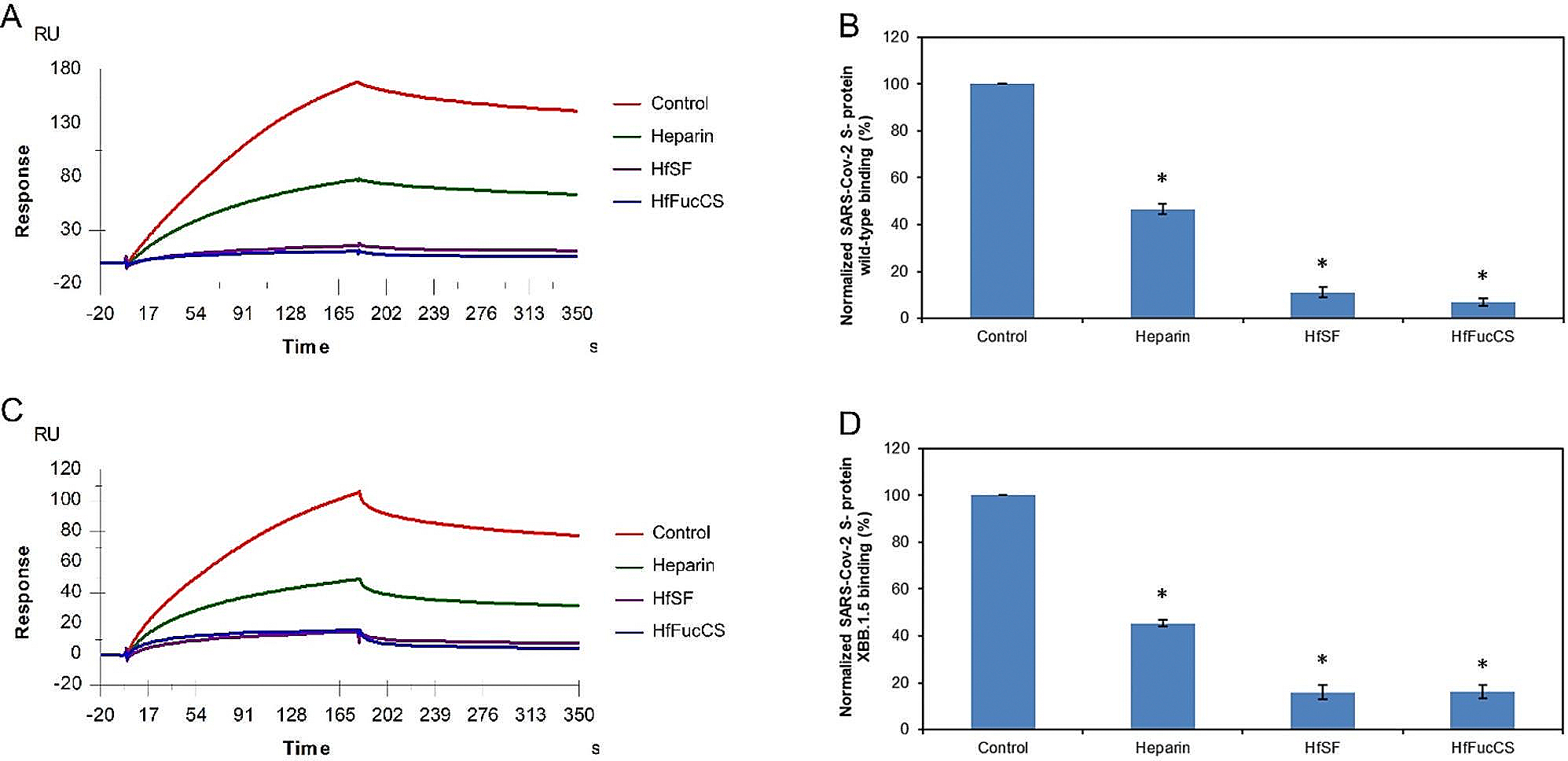
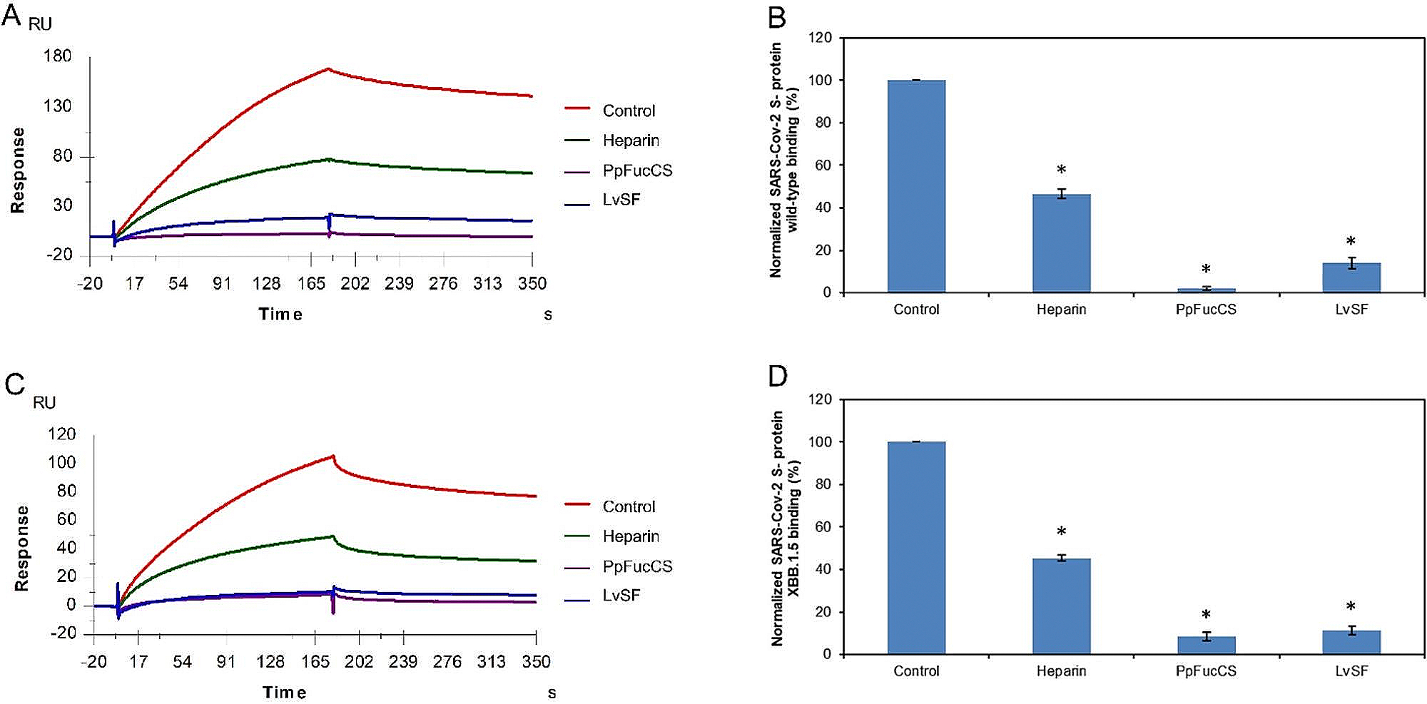
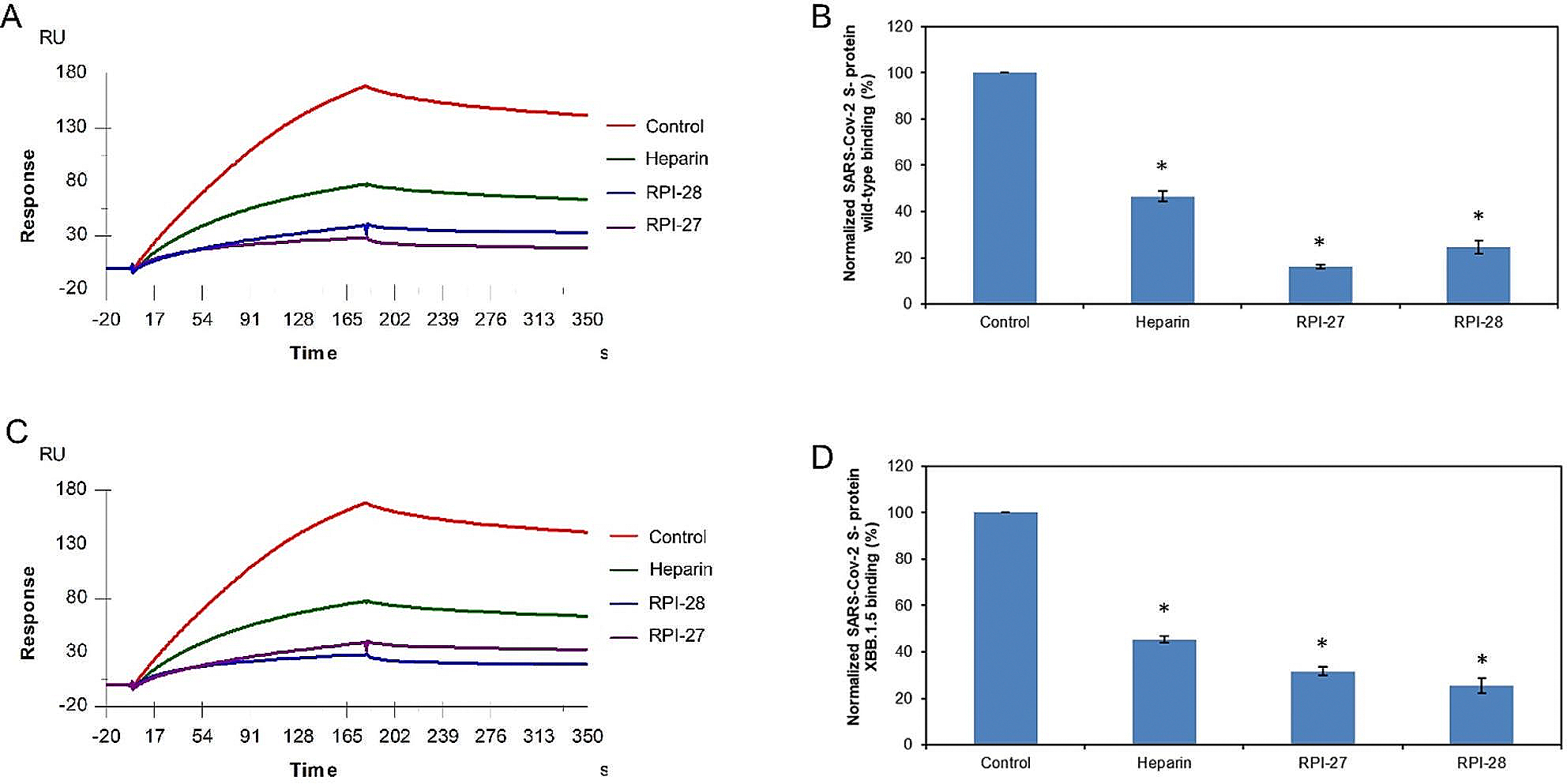
The severe acute respiratory syndrome coronavirus 2 (SARS-CoV-2) has caused a worldwide COVID-19 pandemic, leading to 6.8 million deaths. Numerous variants have emerged since its outbreak, resulting in its significantly enhanced ability to spread among humans. As with many other viruses, SARS‑CoV‑2 utilizes heparan sulfate (HS) glycosaminoglycan (GAG) on the surface of host cells to facilitate viral attachment and initiate cellular entry through the ACE2 receptor. Therefore, interfering with virion-HS interactions represents a promising target to develop broad-spectrum antiviral therapeutics. Sulfated glycans derived from marine organisms have been proven to be exceptional reservoirs of naturally existing HS mimetics, which exhibit remarkable therapeutic properties encompassing antiviral/microbial, antitumor, anticoagulant, and anti-inflammatory activities. In the current study, the interactions between the receptor-binding domain (RBD) of S-protein of SARS-CoV-2 (both WT and XBB.1.5 variants) and heparin were applied to assess the inhibitory activity of 10 marine-sourced glycans including three sulfated fucans, three fucosylated chondroitin sulfates and two fucoidans derived from sea cucumbers, sea urchin and seaweed Saccharina japonica, respectively. The inhibitory activity of these marine derived sulfated glycans on the interactions between RBD of S-protein and heparin was evaluated using Surface Plasmon Resonance (SPR). The RBDs of S-proteins from both Omicrion XBB.1.5 and wild-type (WT) were found to bind to heparin, which is a highly sulfated form of HS. All the tested marine-sourced sulfated glycans exhibited strong inhibition of WT and XBB.1.5 S-protein binding to heparin. We believe the study on the molecular interactions between S-proteins and host cell glycosaminoglycans provides valuable insight for the development of marine-sourced, glycan-based inhibitors as potential anti-SARS-CoV-2 agents.

Keywords: Heparin; Marine sulfated glycans; Omicron XBB.1.5; SARS-CoV-2; SPR; Spike Protein.


Conflict of interest statement

Declarations

Conflict of interest The authors state that they have no conflicts of interest.

Figures

Fig. 1
Fig. 1
Chemical structures of heparin and marine sulfated glycans
Fig. 2
Fig. 2
Omicron phylogenetic tree and S-protein RBD amino acid multiple sequence alignment. (A) Omicron phylogenetic tree, adapted from Nextstrain and CoVariants. (B) Mutation profile of S-protein RBD of WT and XBB.1.5 strains. Multiple sequence alignment was carried out by Clustal Omega (1.2.4). Asterisks (*) indicate positions with a single, fully conserved residue
Fig. 3
Fig. 3
SPR sensorgrams of S-protein RBD of WT and XBB.1.5 binding to heparin. SPR sensorgrams of S-protein RBD binding with heparin; (A) WT and (B) XBB.1.5. Concentrations of S-protein RBD (from top to bottom) are 1000, 500, 250, 125, and 63 nM, respectively
Fig. 4
Fig. 4
Solution competition between heparin and Ib glycans. (A) SPR sensorgrams of the WT SARS-CoV-2 S-protein RBD–heparin interaction competing with different Ib glycans. The concentration of the WT SARS-CoV-2 S-protein RBD was 1 μM mixed with 5 μg/mL of different Ib glycans. (B) Bar graphs (based on triplicate experiments with standard deviation) of normalized WT SARS-CoV-2 S-protein RBD binding preference to surface heparin by competing with different Ib glycans. (C) SPR sensorgrams of the XBB.1.5 SARS-CoV-2 S-protein RBD–heparin interaction competing with different Ib glycans. The concentration of the the XBB.1.5 SARS-CoV-2 S-protein RBD was 1 μM mixed with 5 μg/mL of different Ib glycans. (D) Bar graphs (based on triplicate experiments with standard deviations) of the normalized XBB.1.5 SARS-CoV-2 S-protein RBD binding preference to surface heparin by competing with different Ib glycans. Statistical analysis was performed using an unpaired two-tailed t-test (*: p ≤ 0.05 compared with the control, **: p ≤ 0.01 compared with the control)
Fig. 5
Fig. 5
Solution competition between heparin and Hf glycans. (A) SPR sensorgrams of the WT SARS-CoV-2 S-protein RBD–heparin interaction competing with different Hf glycans. The concentration of the WT SARS-CoV-2 S-protein RBD was 1 μM mixed with 5 μg/mL of different Hf glycans. (B) Bar graphs (based on triplicate experiments with standard deviation) of normalized WT SARS-CoV-2 S-protein RBD binding preference to surface heparin by competing with different Hf glycans. (C) SPR sensorgrams of the XBB.1.5 SARS-CoV-2 S-protein RBD–heparin interaction competing with different Hf glycans. The concentration of the XBB.1.5 SARS-CoV-2 S-protein RBD was 1 μM mixed with 5 μg/mL of different Hf glycans. (D) Bar graphs (based on triplicate experiments with standard deviations) of the normalized XBB.1.5 SARS-CoV-2 S-protein RBD binding preference to surface heparin by competing with different Hf glycans. Statistical analysis was performed using an unpaired two-tailed t-test (*: p ≤ 0.05 compared with the control)
Fig. 6
Fig. 6
Solution competition between heparin and PpFucCS and LvSF. (A) SPR sensorgrams of the WT SARS-CoV-2 S-protein RBD–heparin interaction competing with different PpFucCS and LvSF. The concentration of the WT SARS-CoV-2 S-protein RBD was 1 μM mixed with 5 μg/mL of different PpFucCS and LvSF glycans. (B) Bar graphs (based on triplicate experiments with standard deviation) of normalized WT SARS-CoV-2 S-protein RBD binding preference to surface heparin by competing with different PpFucCS and LvSF glycans. (C) SPR sensorgrams of the XBB.1.5 SARS-CoV-2 S-protein RBD–heparin interaction competing with different PpFucCS and LvSF glycans. The concentration of the the XBB.1.5 SARS-CoV-2 S-protein RBD was 1 μM mixed with 5 μg/mL of different PpFucCS and LvSF glycans. (D) Bar graphs (based on triplicate experiments with standard deviations) of the normalized XBB.1.5 SARS-CoV-2 S-protein RBD binding preference to surface heparin by competing with different PpFucCS and LvSF glycans. Statistical analysis was performed using an unpaired two-tailed t-test (*: p ≤ 0.05 compared with the control)
Fig. 7
Fig. 7
Solution competition between heparin and RPI-27/RPI-28. (A) SPR sensorgrams of the WT SARS-CoV-2 S-protein RBD-heparin interaction competing with RPI-27/RPI-28. The concentration of the WT SARS-CoV-2 S-protein RBD was 1μM mixed with 5 μg/mL of different RPI-27/RPI-28 glycans. (B) Bar graphs (based on triplicate experiments with standard deviation) of normalized WT SARS-CoV-2 S-protein binding preference to surface heparin by competing with different RPI-27/RPI-28 glycans. (C) SPR sensorgrams of the XBB.1.5 SARS-CoV-2 S-protein–heparin interaction competing with different RPI-27/RPI-28 glycans. The concentration of the the XBB.1.5 SARS-CoV-2 S-protein RBD was 1μM mixed with 5 μg/mL of different RPI-27/RPI-28 glycans. (D) Bar graphs (based on triplicate experiments with standard deviations) of the normalized XBB.1.5 SARS-CoV-2 S-protein RBD binding preference to surface heparin by competing with different RPI-27/RPI-28 glycans. Statistical analysis was performed using an unpaired two-tailed t-test (*: p ≤ 0.05 compared with the control)

Similar articles

Cited by

References

    1. Bakhshandeh B, Jahanafrooz Z, Abbasi A, Goli MB, Sadeghi M, Mottaqi MS, Zamani M: Mutations in SARS-CoV-2; consequences in structure, function, and pathogenicity of the virus. Microb. Pathog. 154, 104831 (2021). 10.1016/j.micpath.2021.104831 - DOI - PMC - PubMed
    1. Zhou Y, Zhi H, Teng Y: The outbreak of SARS-CoV-2 omicron lineages, immune escape, and vaccine effectivity. J. Med. Virol. 95 (2023). 10.1002/jmv.28138 e28138. - DOI - PMC - PubMed
    1. Qu P, Faraone JN, Evans JP, Zheng Y-M, Carlin C, Anghelina M, Stevens P, Fernandez S, Jones D, Panchal AR, Saif LJ, Oltz EM, Zhang B, Zhou T, Xu K, Gumina RJ, Liu S-L: Enhanced evasion of neutralizing antibody response by Omicron XBB.1.5, CH.1.1, and CA.3.1 variants. Cell. Rep. 42, 112443 (2023). 10.1016/j.celrep.2023.112443 - DOI - PMC - PubMed
    1. Uriu K, Ito J, Zahradnik J, Fujita S, Kosugi Y, Schreiber G, Sato K: Enhanced transmissibility, infectivity, and immune resistance of the SARS-CoV-2 omicron XBB.1.5 variant. Lancet Infect. Dis. 23, 280–281 (2023). 10.1016/S1473-3099(23)00051-8 - DOI - PMC - PubMed
    1. Gandhi NS, Mancera RL: The structure of glycosaminoglycans and their interactions with proteins. Chem. Biol. Drug Des. 72, 455–482 (2008). 10.1111/j.1747-0285.2008.00741.x - DOI - PubMed

Publication types

MeSH terms

Supplementary concepts