. 2024 Apr 20;14(1):7.
doi: 10.1186/s13395-024-00339-1.

Dimorphic effect of TFE3 in determining mitochondrial and lysosomal content in muscle following denervation

Affiliations

Dimorphic effect of TFE3 in determining mitochondrial and lysosomal content in muscle following denervation

Ashley N Oliveira et al. Skelet Muscle. .

Abstract

Background: Muscle atrophy is a common consequence of the loss of innervation and is accompanied by mitochondrial dysfunction. Mitophagy is the adaptive process through which damaged mitochondria are removed via the lysosomes, which are regulated in part by the transcription factor TFE3. The role of lysosomes and TFE3 are poorly understood in muscle atrophy, and the effect of biological sex is widely underreported.

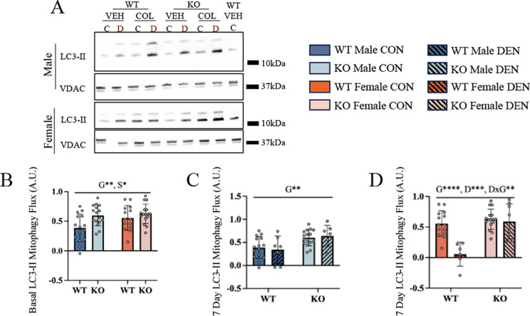
Methods: Wild-type (WT) mice, along with mice lacking TFE3 (KO), a transcriptional regulator of lysosomal and autophagy-related genes, were subjected to unilateral sciatic nerve denervation for up to 7 days, while the contralateral limb was sham-operated and served as an internal control. A subset of animals was treated with colchicine to capture mitophagy flux.

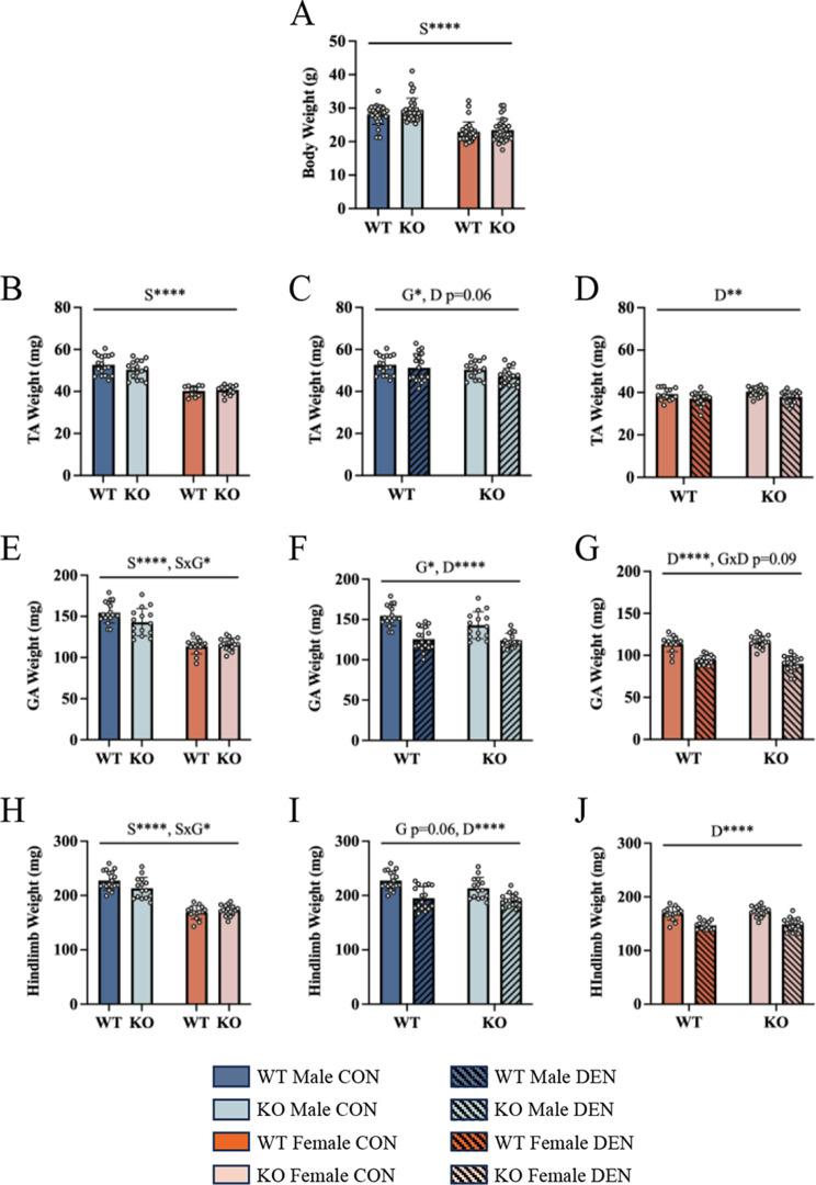
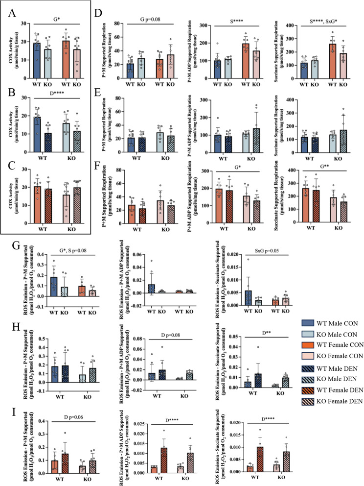
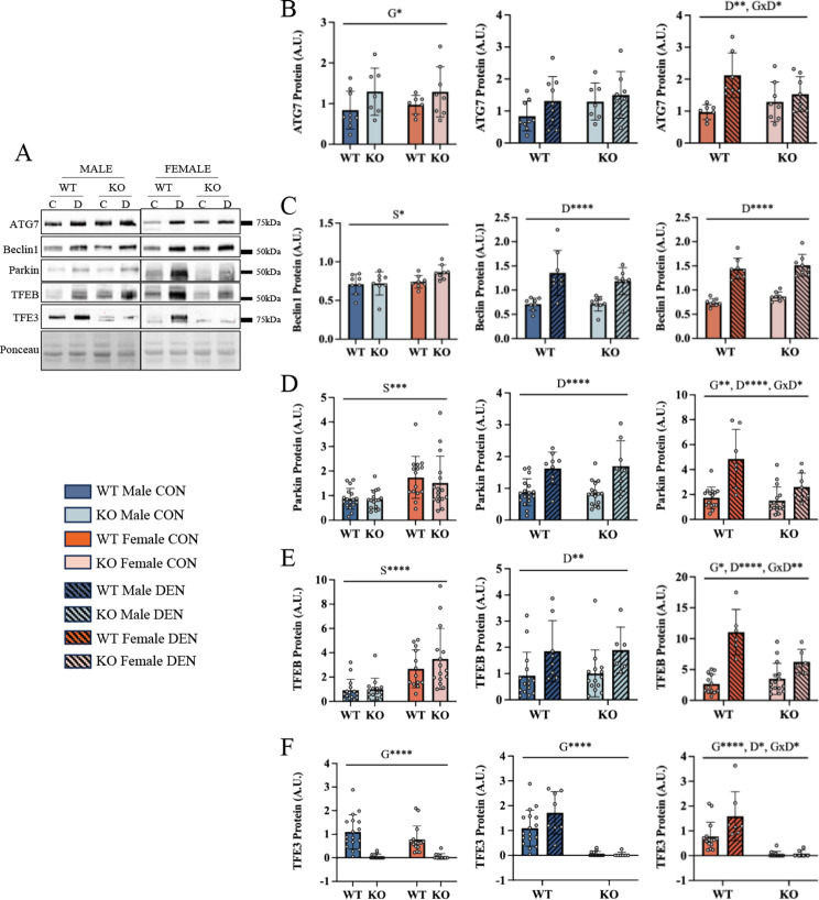
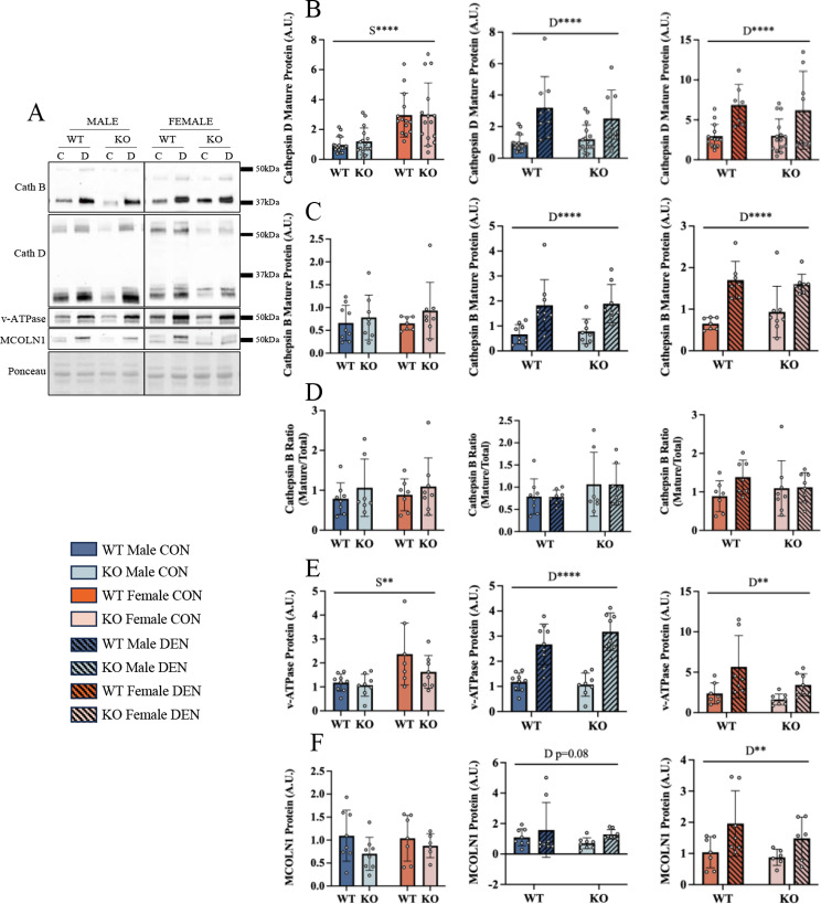
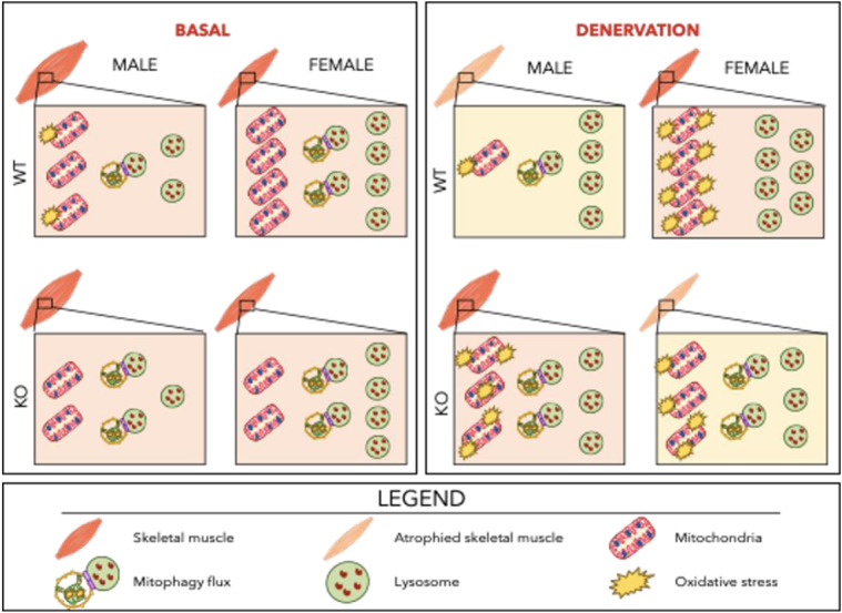
Results: WT females exhibited elevated oxygen consumption rates during active respiratory states compared to males, however this was blunted in the absence of TFE3. Females exhibited higher mitophagy flux rates and greater lysosomal content basally compared to males that was independent of TFE3 expression. Following denervation, female mice exhibited less muscle atrophy compared to male counterparts. Intriguingly, this sex-dependent muscle sparing was lost in the absence of TFE3. Denervation resulted in 45% and 27% losses of mitochondrial content in WT and KO males respectively, however females were completely protected against this decline. Decreases in mitochondrial function were more severe in WT females compared to males following denervation, as ROS emission was 2.4-fold higher. In response to denervation, LC3-II mitophagy flux was reduced by 44% in females, likely contributing to the maintenance of mitochondrial content and elevated ROS emission, however this response was dysregulated in the absence of TFE3. While both males and females exhibited increased lysosomal content following denervation, this response was augmented in females in a TFE3-dependent manner.

Conclusions: Females have higher lysosomal content and mitophagy flux basally compared to males, likely contributing to the improved mitochondrial phenotype. Denervation-induced mitochondrial adaptations were sexually dimorphic, as females preferentially preserve content at the expense of function, while males display a tendency to maintain mitochondrial function. Our data illustrate that TFE3 is vital for the sex-dependent differences in mitochondrial function, and in determining the denervation-induced atrophy phenotype.

Keywords: Autophagy; Lysosomal biogenesis; Mitochondrial respiration; Mitophagy; ROS emission; Sex differences.


Conflict of interest statement

The authors declare no competing interests.

Figures

Fig. 1
Fig. 1
Females are modestly spared from denervation-induced atrophy, as are mice lacking TFE3. TA muscle weights basally stratified by sex (A). TA muscle mass following 7 days of denervation in both WT and KO males (B) and females (C). Gastrocnemius muscle weights basally stratified by sex (D). Gastrocnemius muscle mass following denervation in both WT and KO males (E) and females (F). Sum of hindlimb muscle weights, including TA, EDL, gastrocnemius, and soleus, in WT and KO animals stratified by sex basally (G). Sum of hindlimb muscles weights following denervation in both WT and KO males (H) and females (I). Absolute hindlimb muscle mass lost following denervation (J). Two-way ANOVAs were conducted with repeated measures between CON and DEN conditions, followed by Tukey post-hoc analyses. GA, gastrocnemius; G, denotes a main effect of genotype; S, denotes a main effect of sex; D, indicates a main effect of denervation; SxG, represents an interaction between genotype and sex; GxD, denotes an interaction between genotype and denervation; *, p < 0.05; **, p < 0.01; ****, p < 0.0001; n = 16
Fig. 2
Fig. 2
Sex-dependent response in mitochondrial content and function to denervation. Cytochrome c oxidase (COX) activity assessed as a measure of mitochondrial content basally (A). COX activity following denervation in male animals (B) and female animals (C). Oxygen consumption measured in permeabilized muscle fibers in the presence of pyruvate + malate, ADP and succinate in male and female mice basally (D). Oxygen consumption in the respective states in male mice following denervation (E) and female mice (F). ROS emission rates corrected for respiration supported by pyruvate + malate, ADP, and succinate basally in male and female mice (G). ROS emission rates under the respective states in male mice following denervation. (H) and female mice (I). Two-way ANOVAs were conducted with repeated measures between CON and DEN conditions, followed by Tukey post-hoc analyses. P + M, pyruvate malate; G, represents a main effect of genotype; S, denotes a main effect of sex; D, indicates a main effect of denervation; SxG, represents an interaction between genotype and sex; *, p < 0.05; **, p < 0.01; ****, p < 0.0001; n = 7
Fig. 3
Fig. 3
Females exhibit higher autophagy and mitophagy protein levels and these proteins increase dramatically with denervation in a TFE3-dependent manner. Representative blots for autophagy- and mitophagy-related markers following 7 days (7D) of denervation (A). ATG7 protein content (B) was quantified basally between males and females, then following denervation in male mice and female mice. Similar quantifications were performed for Beclin1 (C), Parkin (D), TFEB (E), and TFE3 (F) protein content. Two-way ANOVAs were conducted with repeated measures between CON and DEN conditions, followed by Tukey post-hoc analyses. G, represents a main effect of genotype; S, denotes a main effect of sex; D, indicates a main effect of denervation; GxS, represents an interaction between genotype and sex; GxS, indicates an interaction effect between genotype and sex; GxS, interaction effect between genotype and sex; *, p < 0.05; **, p < 0.01; ***, p < 0.001; ****, p < 0.0001; control n-14-16, denervated n = 7–8
Fig. 4
Fig. 4
Mitophagic response to chronic denervation is dependent on sex and influenced byTFE3. Representative blots of mitophagy flux following 7 days of denervation (A). Quantification of LC3-II mitophagy flux, taken as the difference between colchicine- and vehicle-treated conditions (A). LC3-II mitophagic flux was assessed basally in male and female mice (B). LC3-II flux was also measured following 7 days of denervation in male mice (C) and female mice (D). Two-way ANOVAs were conducted with repeated measures between CON and DEN conditions, followed by Tukey post-hoc analyses. G, represents a main effect of genotype; S, denotes a main effect of sex; DxG, interaction effect between denervation and genotype; *, p < 0.05; **, p < 0.01; ***, p < 0.001; control n = 14–16, denervated n = 7–8
Fig. 5
Fig. 5
Denervation increases lysosomal markers independent of sex or genotype. Representative blots of lysosomal markers following 7 days of denervation (A). Mature Cathepsin D protein content (B) was quantified basally between males and females, then following denervation in male mice and female mice. Similar quantifications were performed for mature Cathepsin B (C), Cathepsin B ratio (D), v-ATPase (E), and MCOLN1 (F). Two-way ANOVAs were conducted with repeated measures between CON and DEN conditions, followed by Tukey post-hoc analyses. S, denotes a main effect of sex; D, indicates a main effect of denervation; **, p < 0.01; ****, p < 0.0001; control n = 14–16, denervated n = 7–8
Fig. 6
Fig. 6
Summary of findings. Basally, females exhibit increased mitochondrial respiration and modestly reduced ROS emission, and this corresponds with heightened mitochondrial turnover supported by greater lysosomal content and higher basal mitophagy flux in comparison to males. Irrespective of sex, the loss of TFE3 results in a modest loss of mitochondrial content but does not impact lysosomal content. In response to 7 days of denervation, wild type males experience significant muscle atrophy and pronounced declines in mitochondrial content with a modest preservation in function. However, wildtype females appear to preserve mitochondrial content at the expense of function likely due to the reduction in denervation-induced mitophagy despite a robust lysosomal induction. The loss of TFE3 in males appears to shift the denervation-induced response towards that of a wildtype female in that mitochondrial content is modestly preserved at the expense of function. In the females, loss of TFE3 reduces the adaptive lysosomal response to denervation and the inhibitory effect on mitophagy is lost

Similar articles

Cited by

References

    1. Memme JM, Slavin M, Moradi N, Hood DA. Mitochondrial Bioenergetics and Turnover during Chronic Muscle Disuse. Int J Mol Sci. 2021, Vol 22, Page 5179 [Internet]. Multidisciplinary Digital Publishing Institute; 2021 [cited 2022 Jan 10];22:5179. Available from: https://www.mdpi.com/1422-0067/22/10/5179/htm. - PMC - PubMed
    1. Metter EJ, Talbot LA, Schrager M, Conwit R. Skeletal muscle strength as a predictor of all-cause mortality in healthy men. Journals Gerontol - Ser Biol Sci Med Sci. 2002;57:B359–65. doi: 10.1093/gerona/57.10.B359. - DOI - PubMed
    1. Lipes J, Mardini L, Jayaraman D. Sex and mortality of hospitalization admission to an intensive care. Am J Crit Care. 2013;22:314–9. doi: 10.4037/ajcc2013225. - DOI - PubMed
    1. Powers SK, Wiggs MP, Duarte JA, Murat Zergeroglu A, Demirel HA, Zergeroglu AM et al. Mitochondrial signaling contributes to disuse muscle atrophy. Am J Physiol Endocrinol Metab [Internet]. 2012 [cited 2018 Feb 27];303:E31–9. Available from: http://www.pubmedcentral.nih.gov/articlerender.fcgi?artid=3404565%7B&%7D... - PMC - PubMed
    1. Carafoli E, Margreth A, Buffa P. Early biochemical changes in mitochondria from denervated muscle and their relation to the onset of atrophy. Exp Mol Pathol. 1964;3:171–81. doi: 10.1016/0014-4800(64)90050-4. - DOI - PubMed

Substances

Grants and funding