. 2024 Apr 20;14(1):9064.
doi: 10.1038/s41598-024-59110-7.

Epigenetic modulation through BET bromodomain inhibitors as a novel therapeutic strategy for progranulin-deficient frontotemporal dementia

Affiliations

Epigenetic modulation through BET bromodomain inhibitors as a novel therapeutic strategy for progranulin-deficient frontotemporal dementia

Zachary C Rosenthal et al. Sci Rep. .

Abstract

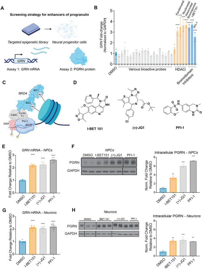
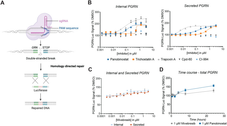
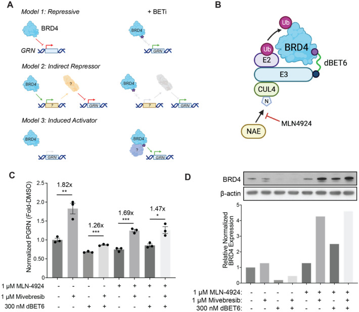
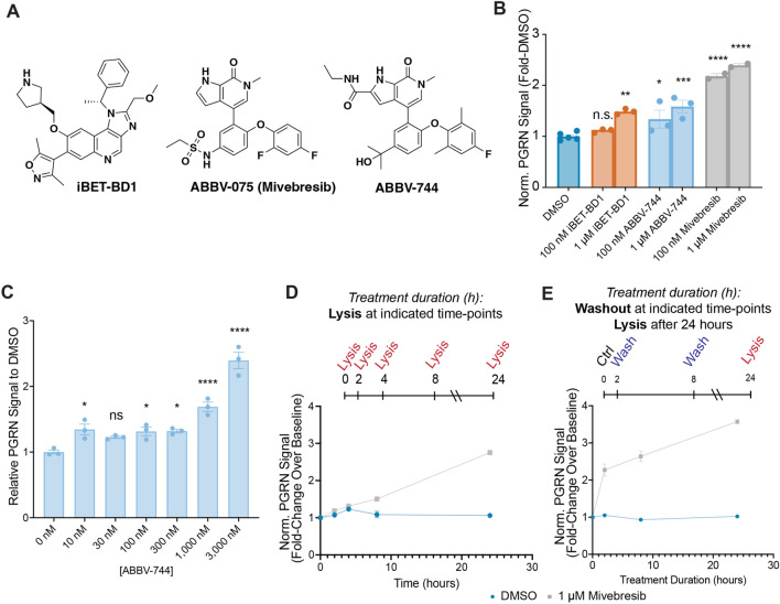
Frontotemporal dementia (FTD) is a debilitating neurodegenerative disorder with currently no disease-modifying treatment options available. Mutations in GRN are one of the most common genetic causes of FTD, near ubiquitously resulting in progranulin (PGRN) haploinsufficiency. Small molecules that can restore PGRN protein to healthy levels in individuals bearing a heterozygous GRN mutation may thus have therapeutic value. Here, we show that epigenetic modulation through bromodomain and extra-terminal domain (BET) inhibitors (BETi) potently enhance PGRN protein levels, both intracellularly and secreted forms, in human central nervous system (CNS)-relevant cell types, including in microglia-like cells. In terms of potential for disease modification, we show BETi treatment effectively restores PGRN levels in neural cells with a GRN mutation known to cause PGRN haploinsufficiency and FTD. We demonstrate that BETi can rapidly and durably enhance PGRN in neural progenitor cells (NPCs) in a manner dependent upon BET protein expression, suggesting a gain-of-function mechanism. We further describe a CNS-optimized BETi chemotype that potently engages endogenous BRD4 and enhances PGRN expression in neuronal cells. Our results reveal a new epigenetic target for treating PGRN-deficient forms of FTD and provide mechanistic insight to aid in translating this discovery into therapeutics.


Conflict of interest statement

S.J.H. serves on the scientific advisory board (SAB) of Proximity Therapeutics, Psy Therapeutics, Frequency Therapeutics, Souvien Therapeutics, Sensorium Therapeutics, 4 M Therapeutics, Ilios Therapeutics, and Entheos Labs, none of whom were involved in the present study. S.J.H. has also received speaking or consulting fees from Amgen, AstraZeneca, Biogen, Merck, Regenacy Pharmaceuticals, Syros Pharmaceuticals, Juvenescence Life, as well as sponsored research or gift funding from AstraZeneca, JW Pharmaceuticals, Lexicon Pharmaceuticals, Vesigen Therapeutics, Compass Pathways, Atai Life Sciences, and Stealth Biotherapeutics. The funders had no role in the design or content of this article, or the decision to submit this review for publication. J.H. is a cofounder of Reelin Therapeutics, which did not provide funding and was not involved in these studies in any way. R.M. is a SAB member and equity holder of Regenacy Pharmaceuticals, ERX Pharmaceuticals, and Frequency Therapeutics. R.M. and N.C.P. are inventors on patent applications related to the CoraFluor TR-FRET probes used in this work. An invention disclosure around novel BET bromodomain compositions and therapeutic uses has been submitted to Massachusetts General Brigham Innovation. All other authors declare no competing interests.

Figures

Figure 1
Figure 1
Bromodomain inhibitors enhance cellular PGRN in human neural progenitor cells. (A) Assay strategy for identifying novel small-molecule enhancers of PGRN. Assay 1: qPCR to identify GRN enhancers. Assay 2: Immunoblotting to validate PGRN enhancement at the protein level. (B) Bromodomain inhibitors (10 µM) enhance GRN mRNA in human NPCs after 24 h. Data are expressed as mean ± S.E. of n = 2–4 compound treatment biological replicates or n = 15 DMSO biological replicates normalized to GAPDH and shown relative to DMSO. Compound concentrations used: bromosporine, 10 µM; I-BET151, 10 µM; PFI-1, 10 µM. (C) Schematic representation of human BRD4 with selected domains and interactors. (D) Structures of BET inhibitor probes used for further investigations. (EF) BET inhibitors enhance GRN mRNA and protein levels in NPCs after 24 h of treatment. (G-H) BET inhibitors enhance PGRN mRNA levels and protein levels in 18-day differentiated neurons after 24 h of treatment. Solid lines on blots indicate discontinuity. Significance determined by Dunnett’s Multiple Comparison, *p < 0.05, **p < 0.01, ***p < 0.001, ****p < 0.0001.
Figure 2
Figure 2
BET inhibitors can enhance lysosomal gene expression and rescue PGRN in GRN-haploinsufficient NPCs and neurons. (A) RNA-seq data of human dermal fibroblasts treated with 1 µM mivebresib for 24 h displaying only lysosomal genes (list from Sardiello et al., Table S1). Significantly altered genes are marked in blue and non-significant changes are marked in orange. Data shown as − log10(Q-value) versus log2(fold-change) of mivebresib treatment compared to DMSO, and represent the average of three biological replicates. (B) Gene Set Enrichment Analysis conducted using lysosomal gene list. (C) Fold-change of GRN as well as selected lysosomal genes shown in (A). (D) Immunocytochemistry of fixed, 2-week growth factor differentiated GRNR493X/+ neurons demonstrating MAP2, neurofilaments (SMI-312R), and TUJ1 expression. (EG) 24 h treatment with mivebresib or panobinostat rescues PGRN haploinsufficiency in GRN-haploinsufficient NPCs and neurons, but not in undifferentiated iPSCs. Significance determined by Dunnett’s Multiple Comparison, *p < 0.05, **p < 0.01, ***p < 0.001, ****p < 0.0001.
Figure 3
Figure 3
BET and HDAC inhibitors can enhance PGRN in the HMC3 PGRN-luciferase reporter line. (A) Schematic for CRISPR-knock in of luciferase at endogenous GRN locus. Adapted from “CRISPR/Cas9 Gene Editing,” by BioRender.com (2024). (B) 20 h treatment with various histone deacetylase inhibitors enhances both internal and secreted PGRN-Luc signal. Data are expressed as mean ± S.E. of 3 treatment biological replicates and 68 DMSO replicates. (C) 18 h treatment with mivebresib enhances both internal and secreted PGRN-Luc signal. Data are expressed as mean ± S.E. of 3 treatment biological replicates and 31 DMSO replicates. (D) Total PGRN-Luc signal after treatment with mivebresib or panobinostat treatment for indicated timeframes. Data are expressed as mean ± S.E. of 2 biological replicates.
Figure 4
Figure 4
Protein dependencies of BET inhibitors as enhancers of PGRN. (A) Mechanistic models for BET inhibition enhancing PGRN protein levels. (B) Mechanism of BRD4 degradation through dBET6. (C) Human NPCs in various co-treatments with MLN-4924 (1 µM), mivebresib (1 µM), and dBET6 (300 nM). Cells were treated with 1 µM MLN-4924 or DMSO only for 2 h prior to the addition of 300 nM dBET6 or DMSO. Cells were then allowed to incubate for four hours prior to the addition of 1 µM mivebresib or DMSO for 24 h. Data are expressed as mean ± S.E. of 3 biological replicates. (D) Immunoblots against BRD4 for human NPCs treated as described in (C), with quantification. Significance determined by Dunnett’s Multiple Comparison, *p < 0.05, **p < 0.01, ***p < 0.001, ****p < 0.0001.
Figure 5
Figure 5
Selectivity and kinetic requirements of BET inhibitors as enhancers of PGRN. (A) Structures of selective BET probes used for these studies. (B) Selective inhibition of BD2 through ABBV-744 (24 h) is sufficient to enhance PGRN protein levels in human NPCs, while pan-BD engagement through mivebresib produces the largest enhancement. Data are expressed as mean ± S.E. of 2–5 biological replicates. For this experiment, one DMSO data point was excluded due to a suspected issue with the total protein assay (value was 40% of the average of the other five data points, Q-value of 0.886 compared to a Qcrit = 0.625 for 6 data points). Significance is compared to the DMSO control group. (C) 24-h treatment with ABBV-744 at various doses enhances PGRN in NPCs. (D) mivebresib (1 µM) potently upregulates PGRN protein levels at the 8-h and 24-h time points in human NPCs. Data are expressed as mean ± S.E. of 2 biological replicates. Significance is compared to the DMSO control group. (E) Treatment with mivebresib for short timeframes, followed by compound washout and then lysis after 24 h from initial compound addition, enhances PGRN protein levels in human NPCs. Data are expressed as mean ± S.E. of 3–6 biological replicates. Significance determined by Dunnett’s Multiple Comparison, *p < 0.05, **p < 0.01, ***p < 0.001, ****p < 0.0001.
Figure 6
Figure 6
Discovery of novel CNS-optimized BET inhibitors as enhancers of PGRN. (A) Optimized poses for ABBV-744 and RAY03-18B docked into BRD4 BDII (PDB: 6ONY). (B) Structures of selected novel BET inhibitors. (C) Novel BET inhibitors (blue) score highly in CNS MPO scoring in comparison to other common BET inhibitors (orange). (D, E) Novel compounds can potently engage BRD4 derived from both HEK-293 T and NPC lysates. Data are expressed as mean ± S.E. of 2 biological replicates. (F, G) Compounds (1 µM) can enhance PGRN in human NPCs after 24 h of treatment. Data are expressed as mean ± S.E. relative to the DMSO control group of 2–3 biological replicates. Data represents compiled experimental runs from different days normalized within each experiment to DMSO. Significance determined by Dunnett’s Multiple Comparison, *p < 0.05, **p < 0.01, ***p < 0.001, ****p < 0.0001.

References

    1. Rademakers R, Neumann M, Mackenzie IR. Advances in understanding the molecular basis of frontotemporal dementia. Nat. Rev. Neurol. 2012;8:423–434. doi: 10.1038/nrneurol.2012.117. - DOI - PMC - PubMed
    1. Rabinovici GD, Miller BL. Frontotemporal lobar degeneration: Epidemiology, pathophysiology, diagnosis and management. CNS Drugs. 2010;24:375–398. doi: 10.2165/11533100-000000000-00000. - DOI - PMC - PubMed
    1. Gass J, Prudencio M, Stetler C, Petrucelli L. Progranulin: An emerging target for FTLD therapies. Brain Res. 2012;1462:118–128. doi: 10.1016/j.brainres.2012.01.047. - DOI - PMC - PubMed
    1. Hosaka T, et al. A novel frameshift GRN mutation results in frontotemporal lobar degeneration with a distinct clinical phenotype in two siblings: Case report and literature review. BMC Neurol. 2017;17:182. doi: 10.1186/s12883-017-0959-2. - DOI - PMC - PubMed
    1. Gass J, et al. Mutations in progranulin are a major cause of ubiquitin-positive frontotemporal lobar degeneration. Hum. Mol. Genet. 2006;15:2988–3001. doi: 10.1093/hmg/ddl241. - DOI - PubMed

MeSH terms