. 2024 Jul;271(7):4336-4347.
doi: 10.1007/s00415-024-12365-6. Epub 2024 Apr 21.

Non-invasive biomarkers for spontaneous intracranial hypotension (SIH) through phase-contrast MRI

Affiliations

Non-invasive biomarkers for spontaneous intracranial hypotension (SIH) through phase-contrast MRI

Katharina Wolf et al. J Neurol. 2024 Jul.

Abstract

Background and objective: Spontaneous intracranial hypotension (SIH) is an underdiagnosed disease. To depict the accurate diagnosis can be demanding; especially the detection of CSF-venous fistulas poses many challenges. Potential dynamic biomarkers have been identified through non-invasive phase-contrast MRI in a limited subset of SIH patients with evidence of spinal longitudinal extradural collection. This study aimed to explore these biomarkers related to spinal cord motion and CSF velocities in a broader SIH cohort.

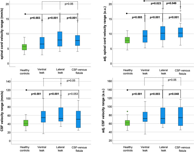
Methods: A retrospective, monocentric pooled-data analysis was conducted of patients suspected to suffer from SIH who underwent phase-contrast MRI for spinal cord and CSF velocity measurements at segment C2/C3 referred to a tertiary center between February 2022 and June 2023. Velocity ranges (mm/s), total displacement (mm), and further derivatives were assessed and compared to data from the database of 70 healthy controls.

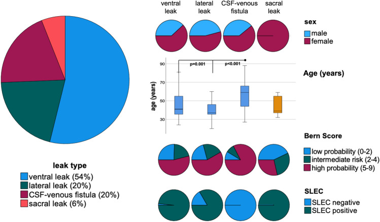
Results: In 117 patients, a leak was located (54% ventral leak, 20% lateral leak, 20% CSF-venous fistulas, 6% sacral leaks). SIH patients showed larger spinal cord and CSF velocities than healthy controls: e.g., velocity range 7.6 ± 3 mm/s vs. 5.6 ± 1.4 mm/s, 56 ± 21 mm/s vs. 42 ± 10 mm/s, p < 0.001, respectively. Patients with lateral leaks and CSF-venous fistulas exhibited an exceptionally heightened level of spinal cord motion (e.g., velocity range 8.4 ± 3.3 mm/s; 8.2 ± 3.1 mm/s vs. 5.6 ± 1.4 mm/s, p < 0.001, respectively).

Conclusion: Phase-contrast MRI might become a valuable tool for SIH diagnosis, especially in patients with CSF-venous fistulas without evidence of spinal extradural fluid collection.

Keywords: CSF flow; CSF–venous fistula; Phase-contrast MRI; Spinal cord motion; Spontaneous intracranial hypotension.


Conflict of interest statement

H. Urbach: honoraria for lectures from Biogen, Eisai, Mbits, Lilly, Co-Editor Clin Neuroradiol. All other authors report no conflict of interest.

Figures

Fig. 1
Fig. 1
Examples of MRI sequences and MRI processing among patients with SIH—top row with sagittal 3D T2 SPACE of the cervical spine, without (middle) and with segmentation (blue = CSF, left, orange = spinal cord, right side). The axial images are phase images with and without automated segmentation. Through-plane axial measurements at C2/C3 (red line) encode velocities in head–feet direction (arrows). At the bottom, acquired velocities in mm/s (y-axis) are plotted over one standardized heartbeat (x-axis). Measurements start with the R-peak of the ECG. Each colored line indicates a different individual. The arrows within the time-resolved plots indicate the peak-to-peak velocity amplitude = velocity range in the craniocaudal direction
Fig. 2
Fig. 2
Clinical data of patients with confirmed spinal CSF leaks, n = 117, spinal longitudinal extradural collection (SLEC). Patients with CSF–venous fistulas were older than patients with ventral or lateral leaks (p < 0.001, p = 0.001, respectively) and showed higher Bern scores (p > 0.082)
Fig. 3
Fig. 3
Boxplots of velocity ranges (top) and adjusted velocity ranges (bottom) in healthy participants (green) and SIH patients (blue) stratified by leak type
Fig. 4
Fig. 4
Receiver operating characteristics of spinal cord motion analysis at the segment C2/C3 in SIH patients with confirmed CSF–venous fistulas. The area under the curve (AUROC) for all spinal cord data is > 0.744, maximum is reached by measurements of the velocity range adjusted to age and sex: 0.83 in patients with CSF–venous fistula

References

    1. Dobrocky T, Nicholson P, Häni L, et al. Spontaneous intracranial hypotension: searching for the CSF leak. Lancet Neurol. 2022;21(4):369–380. doi: 10.1016/S1474-4422(21)00423-3. - DOI - PubMed
    1. Schievink WI, Maya MM, Moser FG, Simon P, Nuño M. Incidence of spontaneous intracranial hypotension in a community: Beverly Hills, California, 2006–2020. Cephalalgia. 2022;42(4–5):312–316. doi: 10.1177/03331024211048510. - DOI - PubMed
    1. Pradeep A, Madhavan AA, Brinjikji W, Cutsforth-Gregory JK. Incidence of spontaneous intracranial hypotension in Olmsted County, Minnesota: 2019–2021. Interv Neuroradiol J Peritherapeutic Neuroradiol Surg Proced Relat Neurosci. 2023 doi: 10.1177/15910199231165429. - DOI - PubMed
    1. Schievink WI, Maya MM, Moser F, Tourje J, Torbati S. Frequency of spontaneous intracranial hypotension in the emergency department. J Headache Pain. 2007;8(6):325–328. doi: 10.1007/s10194-007-0421-8. - DOI - PMC - PubMed
    1. Schievink WI. Spontaneous Spinal Cerebrospinal Fluid Leaks and Intracranial Hypotension. JAMA. 2006;295(19):2286. doi: 10.1001/jama.295.19.2286. - DOI - PubMed

LinkOut - more resources