. 2024 Apr 21;81(1):189.
doi: 10.1007/s00018-024-05215-1.

Adipose-derived stem cells promote glycolysis and peritoneal metastasis via TGF-β1/SMAD3/ANGPTL4 axis in colorectal cancer

Affiliations

Adipose-derived stem cells promote glycolysis and peritoneal metastasis via TGF-β1/SMAD3/ANGPTL4 axis in colorectal cancer

Chaojun Zhu et al. Cell Mol Life Sci. .

Abstract

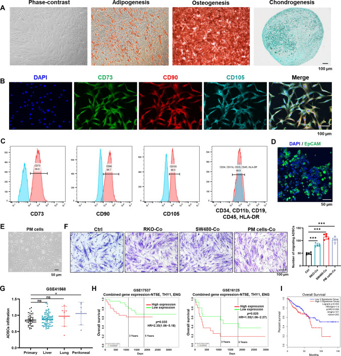
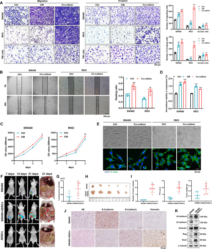
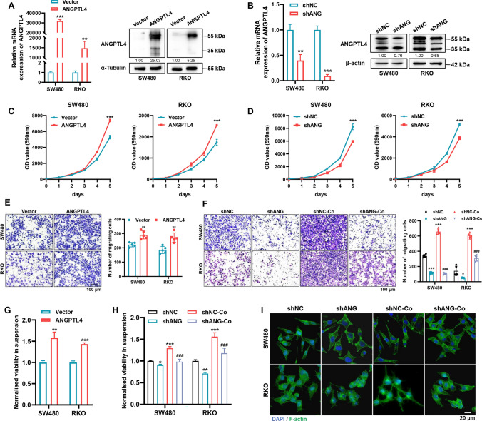
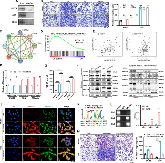
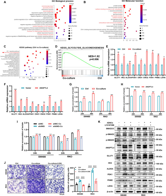
Peritoneal metastasis, the third most common metastasis in colorectal cancer (CRC), has a poor prognosis for the rapid progression and limited therapeutic strategy. However, the molecular characteristics and pathogenesis of CRC peritoneal metastasis are poorly understood. Here, we aimed to elucidate the action and mechanism of adipose-derived stem cells (ADSCs), a prominent component of the peritoneal microenvironment, in CRC peritoneal metastasis formation. Database analysis indicated that ADSCs infiltration was increased in CRC peritoneal metastases, and high expression levels of ADSCs marker genes predicted a poor prognosis. Then we investigated the effect of ADSCs on CRC cells in vitro and in vivo. The results revealed that CRC cells co-cultured with ADSCs exhibited stronger metastatic property and anoikis resistance, and ADSCs boosted the intraperitoneal seeding of CRC cells. Furthermore, RNA sequencing was carried out to identify the key target gene, angiopoietin like 4 (ANGPTL4), which was upregulated in CRC specimens, especially in peritoneal metastases. Mechanistically, TGF-β1 secreted by ADSCs activated SMAD3 in CRC cells, and chromatin immunoprecipitation assay showed that SMAD3 facilitated ANGPTL4 transcription by directly binding to ANGPTL4 promoter. The ANGPTL4 upregulation was essential for ADSCs to promote glycolysis and anoikis resistance in CRC. Importantly, simultaneously targeting TGF-β signaling and ANGPTL4 efficiently reduced intraperitoneal seeding in vivo. In conclusion, this study indicates that tumor-infiltrating ADSCs promote glycolysis and anoikis resistance in CRC cells and ultimately facilitate peritoneal metastasis via the TGF-β1/SMAD3/ANGPTL4 axis. The dual-targeting of TGF-β signaling and ANGPTL4 may be a feasible therapeutic strategy for CRC peritoneal metastasis.

Keywords: ANGPTL4; Adipose-derived mesenchymal stem cells; Colorectal carcinoma; Peritoneal carcinomatosis; TGF-β/SMAD; Warburg effect.


Conflict of interest statement

The authors declare that they have no conflict of interest.

Figures

Fig. 1
Fig. 1
Identification of ADSCs and survival analysis based on ADSCs marker genes. A Representative photomicrographs of ADSCs derived from visceral adipose tissues of CRC patients. The multi-lineage differentiation potential was tested by adipogenesis, osteogenesis, and chondrogenesis. Scale bar 100 µm. B ADSCs were characterized by positive staining for CD73, CD90 and CD105. Scale bar 100 µm. C ADSCs were identified by flow cytometry (blue: isotype control, red: lineage markers). D Immunofluorescence staining of EpCAM, an epithelial tumor marker, in ascites cells. EpCAM epithelial cell adhesion molecule. Scale bar 50 µm. E Third passage PM cells isolated from malignant ascites of CRC patients. Scale bar, 50 µm. F Transwell migration assay of ADSCs co-cultured with indicated CRC cells for 48 h. Scale bar 100 µm. G The infiltration of ADSCs in primary tumors and liver, lung or peritoneal metastases (data from GSE41568). H, I Overall survival plots depicting the survival of CRC patients stratified by the combined gene expression levels of NT5E, THY1, and ENG (data from the PROGgeneV2 and GEPIA2 databases). Data are shown as mean ± SD of at least three independent experiments in (D) (Student’s t test. ns, not significant; *p < 0.05, ***p < 0.001)
Fig. 2
Fig. 2
ADSCs enhance the metastatic potential of CRC cells. Cell migration and invasion assay (A) and wound healing assay (B) in CRC cells treated as indicated. Scale bars 100 µm (A) and 500 µm (B). C Cell proliferation assay on SW480 and RKO cells treated with or without ADSCs-CM. D Anoikis assay results indicating that the viability of tumor cells in suspension was substantially enhanced by treatment with ADSCs-CM or ADSCs for 48 h. E Phalloidin staining showing the morphologic changes and rearrangement of actin filaments in CRC cells. Scale bar 20 µm. F, G Whole-body in vivo imaging analysis of mice (n = 5) intraperitoneally injected with cells as indicated to monitor tumor growth and intraperitoneal dissemination. Image of peritoneal metastases (H) and analyses for tumor number, weight and PCI score (I). IHC staining (J) and western blot analysis (K) for EMT key molecules in intraperitoneal tumors. Scale bar 50 µm. Data are shown as mean ± SD of at least three independent experiments in (AD) (Student’s t test. *p < 0.05, **p < 0.01, ***p < 0.001)
Fig. 3
Fig. 3
ADSCs facilitate the expression of ANGPTL4. A Volcano plot for DEGs in SW480 cells cultured with or without ADSCs. Red: upregulated, green: downregulated, black: not significant; FC: fold change; FDR: false discovery rate. B Venn plot showing three overlapping genes among the DEGs identified by RNA sequencing and those in GSE161097. C ANGPTL4 expression was strikingly increased in SW480 co-cultured with ADSCs for 48 h (n = 3) and in peritoneal metastases compared with primary tumors (GSE161097, n = 15). D qRT-PCR analysis of ANGPTL4 in CRC cells treated as indicated. E Western blot analysis of ANGPTL4 in CRC cells and peritoneal metastasis models. F ANGPTL4 expression levels over clinical stage progression. G Kaplan–Meier of overall survival curve based on ANGPTL4 expression levels (data from GEPIA2). H Multivariate Cox regression analysis of clinical features and ANGPTL4 expression levels (data from TCGA-CRC). I qRT-PCR analysis for ANGPTL4 mRNA expression in CRC cell lines and PM cells. J, K Differential expression of ANGPTL4 in fresh colorectal tumor specimens and adjacent normal tissues of patients from our hospital (n = 62 for qRT-PCR and n = 12 for western blot analysis). Data are shown as mean ± SD of at least three independent experiments in (D) and (I) (Student’s t test. *p < 0.05, **p < 0.01, ***p < 0.001). One-way ANOVA for (F). Paired-sample t test for (J)
Fig. 4
Fig. 4
ANGPTL4 sustains migration capacity and anoikis resistance in CRC cells. A, B qRT-PCR and western blot analysis to detect ANGPTL4 expression level in SW480 and RKO cells transfected with ANGPTL4-overexpression plasmid or ANGPTL4-targeting shRNA. C, D Cell proliferation assay in the indicated CRC cells. E, F Transwell assay results showing that ANGPTL4 modulated the migration ability of CRC cells and ANGPTL4 blockade abolished the promotive effect induced by ADSCs. Scale bar 100 µm. G, H Anoikis resistance was assessed by measuring the viability of cells in suspension. *represents comparison with the shNC group and # represents comparison with the shNC-Co group in (H). I Phalloidin staining showing the morphologic changes and rearrangement of actin filaments in the indicated cells. Scale bar 20 µm. Data are shown as mean ± SD of at least three independent experiments in (AH) (Student’s t test. *p < 0.05, **p < 0.01, ***p < 0.001, ###p < 0.001)
Fig. 5
Fig. 5
ADSCs endow CRC cells with a metastatic phenotype via the TGF-β1/SMAD3/ANGPTL4 axis. A Characterization of exosomes derived from ADSCs by western blot. B Migration assay of CRC cells treated with ADSCs-CM, ADSCs-exosomes, or ADSCs-CM depleted exosomes. Scale bar 100 µm. C PPI analysis between ANGPTL4 and cytokines abundantly secreted by ADSCs. D GSEA of the gene signature associated with the TGF-β signaling pathway in SW480 cells co-cultured with ADSCs. E Correlation of ANGPTL4 with TGFB1 and SMAD3 expression in CRC tissues from GEPIA2. F qRT-PCR analysis for SMAD2 and SMAD3 in indicated CRC cells. G ELISA assay for TGF-β1 in the supernatant of CRC cells, ADSCs and co-culture system. H, I Western blot analysis for key molecules of the TGF-β/SMAD signaling pathway in indicated cells. J Immunofluorescence staining showing the expression and localization of SMAD2/3 (red) and ANGPTL4 (green). Nuclei were counterstained with DAPI (blue). Scale bar 20 µm. K SMAD3 binding sites in the ANGPTL4 promoter, predicted using the JASPAR database. L ChIP assay performed on SW480 cells to confirm the SMAD3 binding sites in the ANGPTL4 promoter. M Migration assay in CRC cells treated as indicated. Cells were treated with or without 10 µM LY2157299 for 48 h. Scale bar 100 µm. Data are shown as mean ± SD of at least three independent experiments in (B, F, GL, M) (Student’s t test. ns, not significant; *p < 0.05, **p < 0.01, ***p < 0.001)
Fig. 6
Fig. 6
ADSCs endow CRC cells with activated glycolysis via the TGF-β1/SMAD3/ANGPTL4 axis. A, B GO enrichment analysis (biologic process and molecular function) of DEGs identified in SW480 cells cultured with or without ADSCs. C KEGG pathway enrichment analysis of DEGs among SW480 cells cultured with or without ADSCs. D GSEA of the gene signature associated with glycolysis and gluconeogenesis in SW480 cells co-cultured with ADSCs. E, F qRT-PCR analysis of the key metabolic enzymes participating in glucose metabolism. GI Quantification of lactate production in CRC cells treated as indicated. *Represents comparison with the shNC group and # represents comparison with the shNC-Co group in (I). J Migration assay in CRC cells treated as indicated. Cells were treated with or without 10 mM 2-DG for 48 h. Scale bar 100 µm. K Western blot analysis results suggesting that treatment with 10 µM LY2157299 for 48 h to inhibit TGF-β/SMAD signaling in CRC cells remarkably reduced the expression of key metabolic enzymes involved in glycolysis. Data are shown as mean ± SD of at least three independent experiments in (EJ) (Student’s t test. ns, not significant; *p < 0.05, **p < 0.01, ***p < 0.001, ###p < 0.001)
Fig. 7
Fig. 7
Dual-targeting of TGF-β signaling and ANGPTL4 strikingly impede the ADSCs-induced intraperitoneal dissemination of CRC cells. A Schematic diagram of CRC intraperitoneal dissemination models showing time points of treatment in mice. B In vivo bioluminescence showing the intraperitoneal tumors in mice treated as indicated. C, D The mice were sacrificed and all the intraperitoneal tumors were collected. EG The number of tumor nodules, PCI score and tumor weight in mice treated as indicated (n = 3). H qRT-PCR analysis of ANGPTL4 in tumors. I Representative images of IHC staining for tumors dissected from mice with corresponding treatments. J Western blot analysis for key molecules involved in TGF-β signaling and glycolysis. Data are shown as mean ± SD in (EH) (Student’s t test. *p < 0.05, **p < 0.01, ***p < 0.001)
Fig. 8
Fig. 8
Targeting TGF-β signaling suppresses the growth of patient-derived organoids sustained by ADSCs. A Phase-contrast images of CRC organoids at 2, 5 and 8 days. Scale bar 100 µm. B, C The viability and number of CRC organoids treated as indicated. D Immunofluorescence staining of SMAD2/3 (red) and ANGPTL4 (green) in CRC organoids. Nuclei were counterstained with DAPI (blue). Scale bar 50 µm. E Schematic illustration showing that ADSCs make CRC cells acquire activated glycolysis and anoikis resistance via the TGF-β1/SMAD3/ANGPTL4 axis, ultimately facilitating peritoneal metastasis and cancer progression. Data are shown as mean ± SD of at least three independent experiments in (B, C) (Student’s t test. **p < 0.01, ***p < 0.001)

Similar articles

Cited by

References

    1. Sung H, Ferlay J, Siegel RL, et al. Global cancer statistics 2020: GLOBOCAN estimates of incidence and mortality worldwide for 36 cancers in 185 countries. CA Cancer J Clin. 2021;71(3):209–249. doi: 10.3322/caac.21660. - DOI - PubMed
    1. Siegel RL, Miller KD, Goding Sauer A, et al. Colorectal cancer statistics. CA Cancer J Clin. 2020;70(3):145–164. doi: 10.3322/caac.21601. - DOI - PubMed
    1. Koppe MJ, Boerman OC, Oyen WJ, et al. Peritoneal carcinomatosis of colorectal origin: incidence and current treatment strategies. Ann Surg. 2006;243(2):212–222. doi: 10.1097/01.sla.0000197702.46394.16. - DOI - PMC - PubMed
    1. Ceelen WP, Bracke ME. Peritoneal minimal residual disease in colorectal cancer: mechanisms, prevention, and treatment. Lancet Oncol. 2009;10(1):72–79. doi: 10.1016/S1470-2045(08)70335-8. - DOI - PubMed
    1. Franko J, Shi Q, Goldman CD, et al. Treatment of colorectal peritoneal carcinomatosis with systemic chemotherapy: a pooled analysis of north central cancer treatment group phase III trials N9741 and N9841. J Clin Oncol. 2012;30(3):263–267. doi: 10.1200/JCO.2011.37.1039. - DOI - PMC - PubMed