Review
. 2024 Apr 22;5(1):14.
doi: 10.1186/s43556-024-00179-x.

NLRP inflammasomes in health and disease

Affiliations
Review

NLRP inflammasomes in health and disease

Zhihao Xu et al. Mol Biomed. .

Abstract

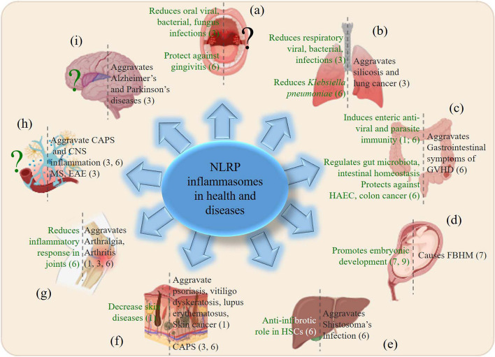
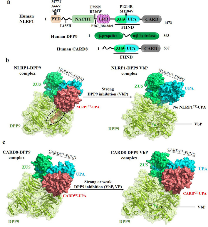
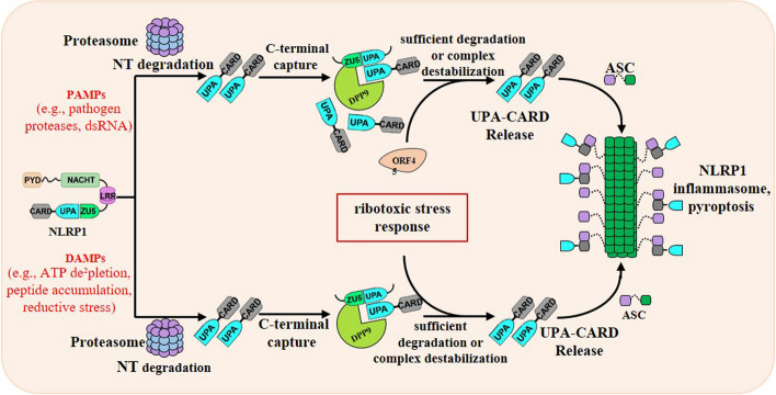
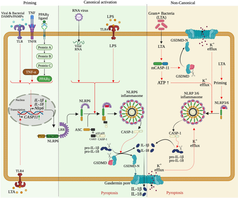
NLRP inflammasomes are a group of cytosolic multiprotein oligomer pattern recognition receptors (PRRs) involved in the recognition of pathogen-associated molecular patterns (PAMPs) and danger-associated molecular patterns (DAMPs) produced by infected cells. They regulate innate immunity by triggering a protective inflammatory response. However, despite their protective role, aberrant NLPR inflammasome activation and gain-of-function mutations in NLRP sensor proteins are involved in occurrence and enhancement of non-communicating autoimmune, auto-inflammatory, and neurodegenerative diseases. In the last few years, significant advances have been achieved in the understanding of the NLRP inflammasome physiological functions and their molecular mechanisms of activation, as well as therapeutics that target NLRP inflammasome activity in inflammatory diseases. Here, we provide the latest research progress on NLRP inflammasomes, including NLRP1, CARD8, NLRP3, NLRP6, NLRP7, NLRP2, NLRP9, NLRP10, and NLRP12 regarding their structural and assembling features, signaling transduction and molecular activation mechanisms. Importantly, we highlight the mechanisms associated with NLRP inflammasome dysregulation involved in numerous human auto-inflammatory, autoimmune, and neurodegenerative diseases. Overall, we summarize the latest discoveries in NLRP biology, their forming inflammasomes, and their role in health and diseases, and provide therapeutic strategies and perspectives for future studies about NLRP inflammasomes.

Keywords: Auto-inflammatory; Autoimmune; Health and disease; NLRP inflammasome; Neurological disorders; Therapeutic inhibitor.


Conflict of interest statement

The authors declare that they have no competing interests.

Figures

Fig. 1
Fig. 1
Overview of Structural organization of NLRP family and NLRP inflammasome assembly. NLRP family genes consist of an N-terminal pyrin domain (PYD), a central nucleotide binding and oligomerization (NACHT) domain, bounded by C-terminal leucine-rich repeats (LRRs) and caspase recruitment (CARD) or pyrin (PYD) domains. In NLRP family, NLRP1 (and its derived CARD8) sensors alone contain a FIIND domain and a CARD domain a C-terminal, NLRP10 sensor alone only consists of a PYD and a NACHT domains, while the remaining are similar, but activated by different stimuli. Numerous NLRP stimuli can trigger NLRP activation to an assembled inflammasome, and these stimuli include microbe-derived signals (commensal bacteria and commensal fungi), pathogen-derived signals (foreign bacteria, fungi, parasites, and viruses), and host-derived signals (ion flux, mitochondrial dysfunction and damages, ROS, and metabolic factors). A bona fide assembled NLRP inflammasome consists of a cytosolic a NLRP gene (the sensor), an apoptosis-associated speck-like (ASC) protein containing a caspase activation and recruitment domain, CARD (the adaptor), and a cysteine protease caspase-1 (CASP1) (the effector)
Fig. 2
Fig. 2
Roles of NLRP in health and diseases in different organs and tissues. Schematic representation of the different functions of NLRP in (a) oral cavity, (b) lungs, (c) digestive system, (d) pregnancy and fetus, (e) liver, (f) skin, (g) joints, (h) peripheral and central nervous system, (i) brain. Functions shown in green represent the NLRP inflammasome-dependent protective response (or NLRP beneficial roles in health) and functions shown in black represent the NLRP inflammasomes-associated diseases (or NLRP detrimental roles in diseases). Numbers in brackets indicate the different NLRPs associated. “?” indicates unknown roles of NLRP inflammasomes
Fig. 3
Fig. 3
Structural features of human NLRP1. a Domain organization of NLRP1, DPP9 and CARD8. Human NLRP1 and CARD8 autoproteolysis sites are shown between the ZU5 and UPA subdomains of the FIIND. The dotted black circle shows the N-terminus of NLRP1CT-UPA folded into the DPP9 active-site tunnel. b Cryo-EM structure of the hNLRP1FL-DPP9-hNLRP1CT ternary complex. NLRP1 directly bonds to the DPP9 active site and strong DPP9 inhibitors (e.g., VbP) are required to displace the inserted peptide of hNLRP1CT in the DPP9 catalytic pocket and destabilize the ternary complex. c Cryo-EM structure of the hCARD8FL-DPP9-hCARD8CT ternary complex. Unlike human NLRP1, the precisely mechanism of the ternary complex destabilized by DPP9 inhibitors (e.g., VbP, VP) remains unknown because CARD8 does not directly bond to the DPP9 active site
Fig. 4
Fig. 4
The mechanisms of human NLRP1 inflammasome activation by PAMPs and DAMPs. The proteasome-mediated degradation of N-terminus of NLRP1 liberates the C-terminal UPA-CARD from autoinhibition. The acceleration of degradation of NLRP1 by several PAMPs (for example, pathogen proteases) overwhelms the DPP9 ternary complex checkpoint to oligomerize into an inflammasome. On the other hand, the destabilization of the DPP9 ternary complex by several DAMPs (for example, peptide accumulation) releases the C-terminal functional domain of NLRP1 to assemble into an inflammasome. There are two distinct signals-sufficient degradation and repressive complex destabilization-create an unstable state to active NLRP1 inflammasome
Fig. 5
Fig. 5
The mechanisms of human NLRP3 inflammasome activation and regulation. The activation of NLRP3 inflammasome occurs either through a canonical two-step pathway or a non-canonical pathway, and a direct or alternative pathway. The canonical activation pathway involved 2 steps: a priming (signal 1, left panel) and an activation (signal 2, second panel from left) steps. Priming step is induced by NLRP3 signals, including LPS and TNF, IL-1b, IFNs, lipopolysaccharide (LPS), and sphingosine-1 phosphate (S1P), activate NF-κB that; in turn upregulates the transcription of Nlrp3 gene and other genes (ASC and pro-caspase1) involved in NLRP inflammasome, by interacting with and triggering their receptors. Once transcribed, NLRP3 is pre-activated by interacting with NEK7, forming a complex that will be activated into hetero-complex inflammasome. The canonical activation of NLRP3 inflammasome is induced by signal 2 including PAMPs (nigericin, viral RNA, and MDP) and DAMPs (extracellular ATP, mtDNA, and mtROS) and particulates. The molecular mechanisms behind the polymerization and the activation of NLRP3 inflammasome include activation of several signaling events, including induction of K+ efflux, Ca2+ flux, Clefflux, lysosomal disruption, mtROS production, and release of oxidized mtDNA in the cytosolic compartment. Thus, formation of NLRP3 inflammasome includes oligomerization of NLRP3-NEK7, recruitment of ASC, and Casp1. auto-proteolysis of proteolytic cleavage of Casp1 releases p10/p20 active enzyme, which digest Pro-IL-1β and Pro-IL-18 into IL-1β and IL-18 cytokines to promote proinflammatory responses. The subunit p10/p20 of Casp1 also digests GSDMD releasing GSDMD-N that form cell membrane pore to result in pyroptosis of the cell. Non-canonical activation of NLRP3 inflammasome (third panel from left) occurs without priming, as Casp4 is already present in the cytoplasm, and is induced by gram-negative bacteria that release LPS into the cell cytosol. Released LPS activates Casp11 in human (and Casp4/5 in mouse), which cleaves GSDMD complex releasing GSDMD-N that forms gasdermin pores and induces pyroptosis. The gasdermin pore formed constitutes a channel for K+ efflux, which activates the NLRP3 inflammasome, and consequently activate Casp1 and IL-1β and IL-18. The alternative pathway (right panel) activation is induced by TLR4 agonists that activates the TLR4-TRIF-RIPK1-FADD-Casp8 signaling pathway. Consequently, Casp8 activates the NLRP3 inflammasome. Note that, there is no need of K+ efflux, ASC speck formation, to activate inflammasome, and there is no pyroptosis
Fig. 6
Fig. 6
The mechanisms of human NLRP6 inflammasome activation. The activation of NLRP6 inflammasome obey a two-steps mechanism: a priming and an activation. In the priming step, induction of the Nlrp6 gene transcription and other NLRP6 inflammasome components is triggered by TNF‐α, viral and bacterial PAMPs/DAMPs, and/or the peroxisome proliferator‐activated receptor-γ (PPAR‐γ). Once translated, NLRP6 inflammasome is activated by dsARN from RNA virus and LPS, and occurs through NLRP6 recruitment of ASC and Casp1. The activated NLRP6 inflammasome activates IL-1β, and IL-18 from their respective precursors (pro-IL-1β, and pro-IL-18, respectively) by catalytic cleavage. NLRP6 inflammasome also catalyses digestion of GSDMD into GSDMD-C and GSDMD-N that forms gasdermin membrane pore, which yields to pyroptosis. IL-1β and IL-18 cytokines are release out of the cells to promote pro-inflammatory responses. Besides, the NLRP6 protein is also found in the cytosol in low level and is autoinhibited in quiescent cell condition. In this condition, LTA from Gram + bacteria induce an indirect non-canonical activation of the NLRP6 inflammasome. LTA activates caspase-11 (involved in the non-canonical inflammasome activation pathway) which trigger activation of NLRP3/6 inflammasomes, through production of ions flux (specifically K+ efflux via GSDMD pores), which in turn activate caspase-1 and release IL-1β and IL-18

Similar articles

Cited by

References

    1. Janeway, C.A., Jr. & Medzhitov, R. Innate immune recognition. Annu Rev Immunol. 2002;20197–216. 10.1146/annurev.immunol.20.083001.084359. - PubMed
    1. Leal, V.N.C. & Pontillo, A. Canonical Inflammasomes. Methods Mol Biol. 2023;26961–27. 10.1007/978-1-0716-3350-2_1. - PubMed
    1. Schroder K, Tschopp J. The inflammasomes. Cell. 2010;140(6):821–832. doi: 10.1016/j.cell.2010.01.040. - DOI - PubMed
    1. Medzhitov R. Recognition of microorganisms and activation of the immune response. Nature. 2007;449(7164):819–826. doi: 10.1038/nature06246. - DOI - PubMed
    1. Martinon F, Burns K, Tschopp J. The inflammasome: a molecular platform triggering activation of inflammatory caspases and processing of proIL-beta. Mol Cell. 2002;10(2):417–426. doi: 10.1016/s1097-2765(02)00599-3. - DOI - PubMed

Publication types

MeSH terms

LinkOut - more resources