. 2024 Oct;112(10):1803-1816.
doi: 10.1002/jbm.a.37725. Epub 2024 Apr 21.

Injectable bioactive poly(propylene fumarate) and polycaprolactone based click chemistry bone cement for spinal fusion in rabbits

Affiliations

Injectable bioactive poly(propylene fumarate) and polycaprolactone based click chemistry bone cement for spinal fusion in rabbits

Xifeng Liu et al. J Biomed Mater Res A. 2024 Oct.

Abstract

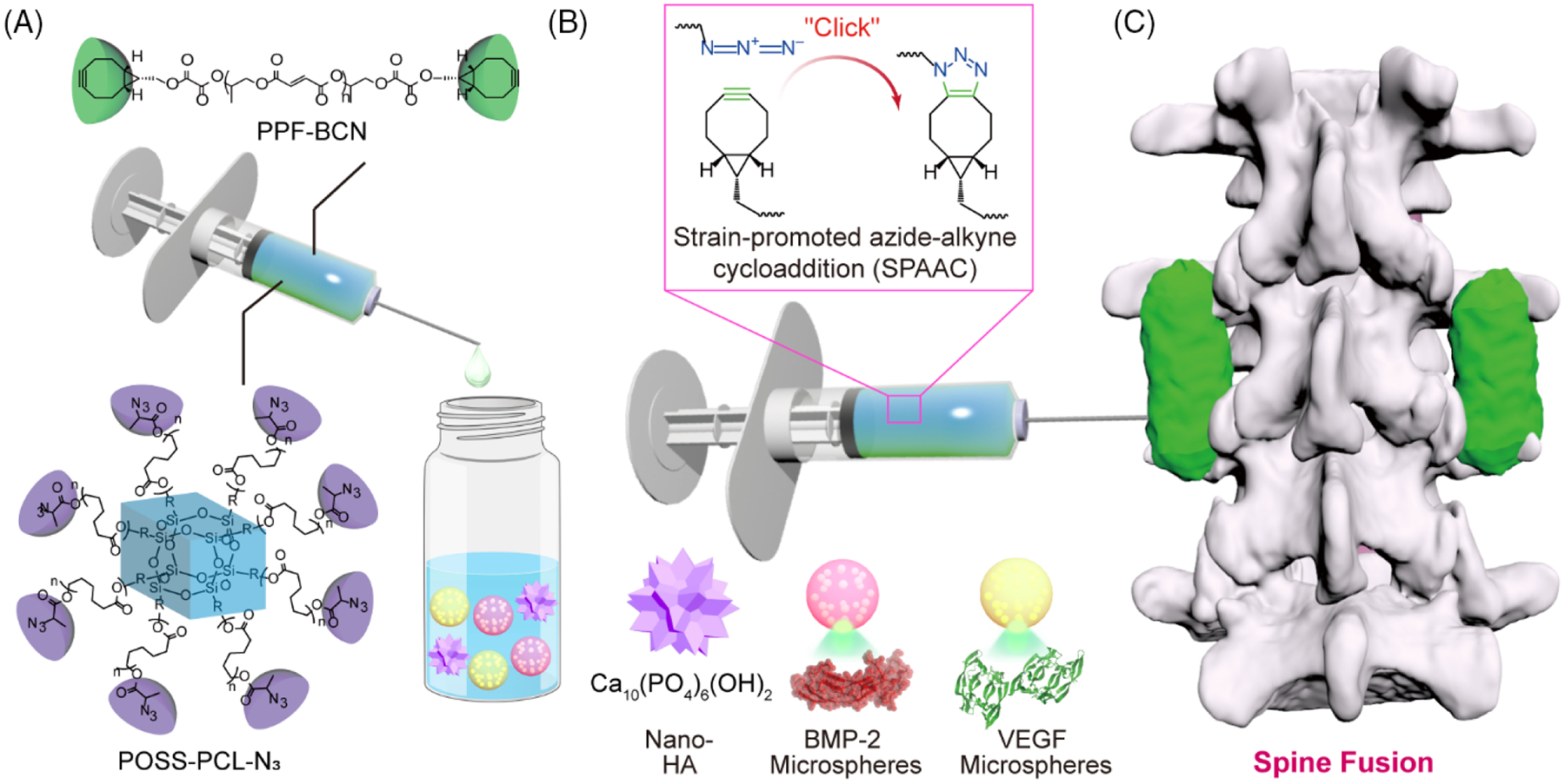
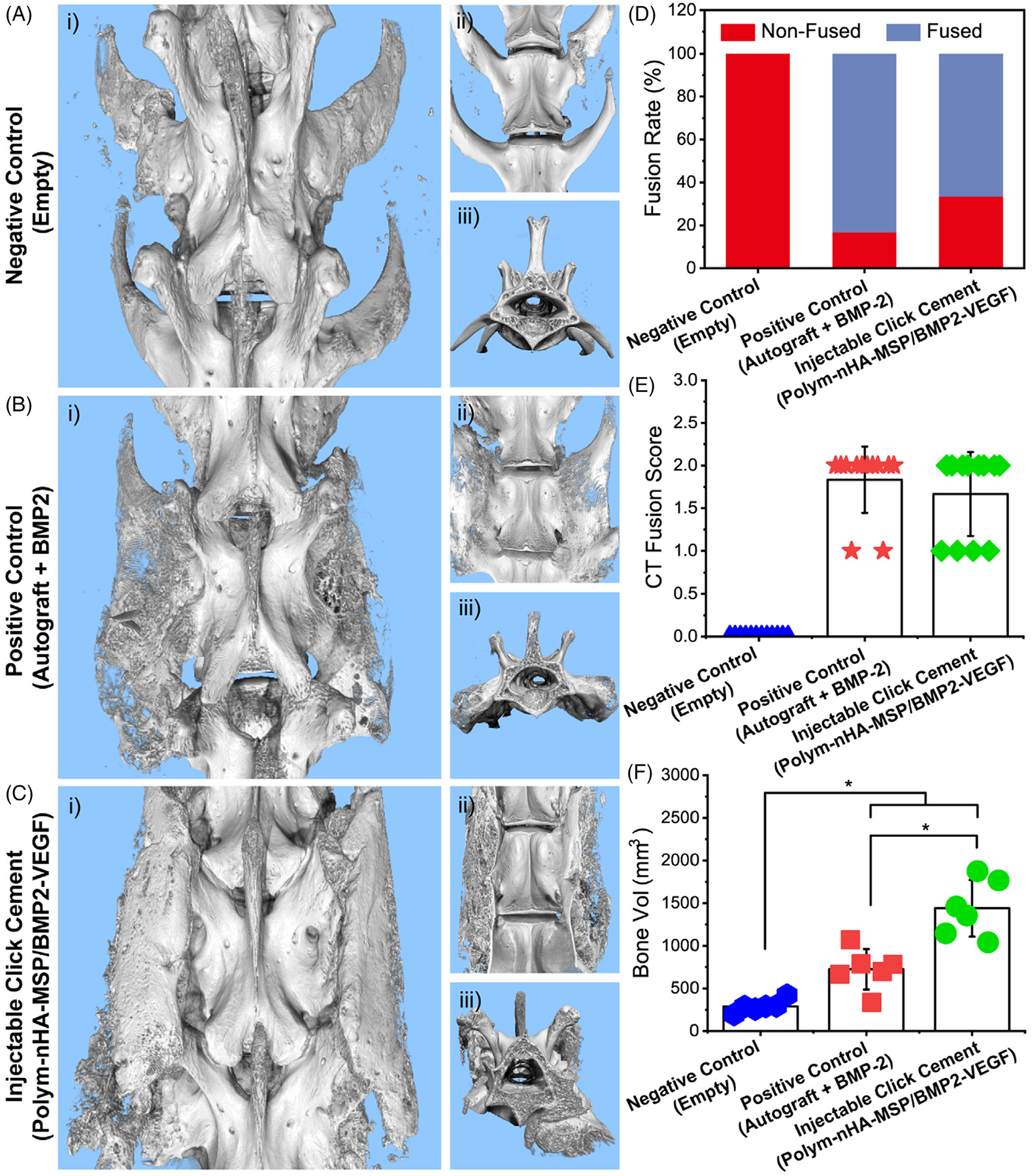
Degenerative spinal pathology is a widespread medical issue, and spine fusion surgeries are frequently performed. In this study, we fabricated an injectable bioactive click chemistry polymer cement for use in spinal fusion and bone regrowth. Taking advantages of the bioorthogonal click reaction, this cement can be crosslinked by itself eliminating the addition of a toxic initiator or catalyst, nor any external energy sources like UV light or heat. Furthermore, nano-hydroxyapatite (nHA) and microspheres carrying recombinant human bone morphogenetic protein-2 (rhBMP-2) and recombinant human vascular endothelial growth factor (rhVEGF) were used to make the cement bioactive for vascular induction and osteointegration. After implantation into a rabbit posterolateral spinal fusion (PLF) model, the cement showed excellent induction of new bone formation and bridging bone, achieving results comparable to autograft control. This is largely due to the osteogenic properties of nano-hydroxyapatite (nHA) and the released rhBMP-2 and rhVEGF growth factors. Since the availability of autograft sources is limited in clinical settings, this injectable bioactive click chemistry cement may be a promising alternative for spine fusion applications in addressing various spinal conditions.

Keywords: bioorthogonal click chemistry; bone cement; injectable polymers; spine fusion; tissue engineering.


Conflict of interest statement

Dr. Elder is a consultant for DePuy Synthes and SI bone, owns stock options and is on the medical advisory board for Injectsense, and receives institutional support for clinical trials from Stryker and SI Bone. The other authors declare no competing financial interest.

Figures

FIGURE 1
FIGURE 1
(A) The fabrication of injectable bioactive cement containing two predissolved polymer compositions with nano-HA and microspheres loaded with rhBMP-2 or rhVEGF growth factors. (B) Crosslinking of polymer systems via biorthogonal click chemistry. (C) Enhanced bone generation, taking advantage of bridging the bone gap with click cement, and vascularization and osteointegration from rhBMP-2 and rhVEGF growth factors.
FIGURE 2
FIGURE 2
(A) SEM of the dried microspheres. Photographs of the fabricated click chemistry cement as injected with (B) small 1 mL syringes and (C) large 10 mL syringes. (D) SEM images of the dried click chemistry cement with (E) a detailed view of the microspheres embedded within. (F) Pores were generated due to potential swelling, shrinking, and degradation of microspheres. (G) A typical stress–strain curve tested for the click chemistry bone cement with averaged compressive modulus (n = 3). The release kinetics of (H) BMP-2 and (I) VEGF growth factors from the microspheres (n = 3).
FIGURE 3
FIGURE 3
The in vitro cell studies. (A) Viabilities of rBMSC and HUVEC after coculture with the click cement (n = 3). The proliferation of (B) rBMSC and (C) HUVEC cells on the crosslinked click chemistry bone cement with or without rhBMP-2/rhVEGF growth factors (nHA: nano-hydroxyapatite; MSP: microspheres; n = 3). (D) Osteogenic differentiation of rBMSCs on the click chemistry bone cement with or without incorporation of growth factors within the embedded microspheres by immunofluorescence staining of osteogenic ALP marker. (E) Vascular differentiation of HUVEC cells on the cement with or without incorporation of growth factors by immunofluorescence staining of CD31 vascular marker. *p < 0.05.
FIGURE 4
FIGURE 4
Animal surgery and characterization. (A) Schematic demonstration of the implantation of click chemistry bone cement in rabbits to mimic human spine fusion as demonstrated using the human posterolateral spinal fusion (PLF) model. (B) Photograph of the surgical process in the operation room. (C) X-ray images of the rabbit spine section for the empty negative control, autograft positive control, and injectable bioactive click chemistry cement group.
FIGURE 5
FIGURE 5
Micro-CT images of the rabbit spine section for the (A) empty negative control, (B) autograft positive control group, and (C) injectable bioactive click chemistry cement (i: posterior view; ii: interior view; iii: vertical view). (D) Fusion rate percentage for different groups with varied fusion status (non-fused or fused). (E) Fusion score evaluated from CT images with 0 = nonfusion, 1 = partial fusion, and 2 = fused (n = 12: averaged from two surgeons independently scored for six spine implantation sites, total 12 scores per group) for the empty negative control, autograft positive control, and injectable bioactive click cement. (F) Bone volume calculated for the empty negative control, autograft positive control, and injectable bioactive click cement (n = 6 spine implantation sites). *p < 0.05.
FIGURE 6
FIGURE 6
The sliced views from the coronal plane, sagittal plane, and axial plane for the (A) empty negative control, (B) autograft positive control group, and (C) injectable bioactive click chemistry cement. Arrows indicate the new bone development in the spine fusion site. H & E staining histological analysis of rabbit spine section of (D) empty negative control, (E) autograft positive control, and (F) injectable bioactive click chemistry cement after slicing (*indicates original bone; arrow indicates newly regenerated bone).

References

    1. Veronesi F, Borsari V, Martini L, et al. The impact of frailty on spine surgery: systematic review on 10 years clinical studies. Aging Dis. 2021;12(2):625–645. - PMC - PubMed
    1. Parenteau CS, Lau EC, Campbell IC, Courtney A. Prevalence of spine degeneration diagnosis by type, age, gender, and obesity using Medicare data. Sci Rep. 2021;11(1):5389. - PMC - PubMed
    1. Diebo BG, Shah NV, Boachie-Adjei O, et al. Adult spinal deformity. The Lancet. 2019;394(10193):160–172. - PubMed
    1. Martin BI, Mirza SK, Spina N, Spiker WR, Lawrence B, Brodke DS. Trends in lumbar fusion procedure rates and associated hospital costs for degenerative spinal diseases in the United States, 2004 to 2015. Spine. 2019;44(5):369–376. - PubMed
    1. Kamson S, Lu D, Sampson PD, Zhang Y. Full-endoscopic lumbar fusion outcomes in patients with minimal deformities: a retrospective study of data collected between 2011 and 2015. Pain Physician. 2019;22(1):75–88. - PubMed

Publication types

MeSH terms

LinkOut - more resources