Review
. 2024 Dec;8(12):e2400058.
doi: 10.1002/smtd.202400058. Epub 2024 Apr 21.

Characterizing Prion-Like Protein Aggregation: Emerging Nanopore-Based Approaches

Affiliations
Review

Characterizing Prion-Like Protein Aggregation: Emerging Nanopore-Based Approaches

Nathan Meyer et al. Small Methods. 2024 Dec.

Abstract

Prion-like protein aggregation is characteristic of numerous neurodegenerative diseases, such as Alzheimer's and Parkinson's diseases. This process involves the formation of aggregates ranging from small and potentially neurotoxic oligomers to highly structured self-propagating amyloid fibrils. Various approaches are used to study protein aggregation, but they do not always provide continuous information on the polymorphic, transient, and heterogeneous species formed. This review provides an updated state-of-the-art approach to the detection and characterization of a wide range of protein aggregates using nanopore technology. For each type of nanopore, biological, solid-state polymer, and nanopipette, discuss the main achievements for the detection of protein aggregates as well as the significant contributions to the understanding of protein aggregation and diagnostics.

Keywords: amyloid fibers; nanopore; prion‐like proteins oligomers; single‐molecule techniques.


Conflict of interest statement

The authors declare no conflict of interest.

Figures

Figure 1
Figure 1
Diagram of amyloid protein aggregation. The kinetics follow a sigmoidal pattern, which can be broken down into 3 phases (lag, exponential, plateau). The species present in the lag phase, mainly oligomers, are insensitive to Thioflavin T, as they are not structured into β‐sheets. At the end of the aggregation process (plateau), the fibers are organized into plaques, which are responsible for various neurodegenerative diseases.
Figure 2
Figure 2
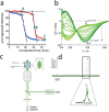
a) Intensity of the peak corresponding to the amide and aromatic regions monitored by NMR as a function of time. With time, the intensity decreases, indicating aggregation. The blue and red curves correspond to aggregation of IAPP (amyloid protein) in the absence and presence of curcumin respectively. Figure adapted from.[ 11a ] Copyright 2012, American Chemical Society b) Superimposed DC spectra recorded at 45‐min time intervals. The increase in the amount of amyloid−β is indicated by the increase in a negative band at 215 nm and a positive band at 195 nm. Figure adapted from.[ 16b ] Copyright 2007, Wiley c) Experimental diagram of a device for detecting amyloid aggregates by confocal spectroscopy. d) fluorescence photons as a function of time measured, when a ThT‐bound aggregate passes through the volume, a burst of fluorescence is measured. The properties of this burst, in particular its duration, provide information on the properties of the aggregate that has passed through the confocal volume (its diffusion coefficient, friction coefficient, etc.).
Figure 3
Figure 3
Left, experimental principle of the resistance pulse. Right, schematic representation of current blockage when an analyte passes through the pore. The current I0 corresponds to the open pore, and ΔI corresponds to the current blockage when a molecule transiently blocks the pore. Δt corresponds to the duration of current blockage.
Figure 4
Figure 4
a) Example of different biological nanopores. Figure adapted from.[ 28a ] Copyright 2018, Wiley b) Detection of the different species formed during the aggregation of Αβ with an a‐hemolysin nanopore. Figure adapted from.[ 40 ] Copyright 2021, Elsevier c) Detection and discrimination of Αβ 1‐42 peptides varying by only one amino acid in their sequence by an aerolysin nanopore. Figure adapted from.[ 51 ] Copyright 2022, Wiley.
Figure 5
Figure 5
a) Schematic of a SiN nanopore with a low aspect ratio. b) Detection of the different aggregates formed during aggregation of the Αβ 1‐42 peptide by SiN nanopore. With increasing incubation time, blocking events have larger amplitudes and longer timescales, suggesting the formation of increasingly large aggregates. Figure adapted from.[ 94 ] Copyright, 2012, American Chemical Society c) Estimated volumes of oligomers formed following α−synuclein aggregation from measured ΔI/I blocking events. Estimated volumes correlate well with microscopic characterization techniques. Figure adapted from.[ 58 ] Copyright 2023, American Chemical Society.
Figure 6
Figure 6
a) Schematic of a nanopipette with a high aspect ratio. b) Detection of aggregation seeding by a small amount of aggregation by RT‐FaST. The control (blue) shows no blocking events, while the seeded condition (red) does. Figure adapted from.[ 76 ] Copyright, 2023, American Chemical Society c) Increase in signal‐to‐noise ratio by a factor of 6 after addition of polyethylene glycol to clog the system. PEG clogging enabled detection and discrimination of unfragmented and fragmented α−synuclein fibers. Figure adapted from.[ 75 ] Copyright, 2020, American Chemical Society.
Figure 7
Figure 7
a) Schematic diagram of the different geometries that can be obtained with etched trace nanopore technology, depending on the experimental aperture conditions. b) Left, aggregation kinetics monitored by ThT in the absence (black) and presence (red) of pyrimethanil. Samples formed during the early phase (circled in dotted black) were analyzed by conical etched trace nanopore (right of Figure). Figure adapted from.[ 89 ] Copyright, 2021, Elsevier.

References

    1. Ross C. A., Poirier M. A., Nat. Med. 2004, 10, S10. - PubMed
    1. a) Morris A. M., Watzky M. A., Finke R. G., Biochim. Biophys. Acta 2009, 1794, 375; - PubMed
    2. b) Linse S., Pure Appl. Chem. 2019, 91, 211;
    3. c) Sakono M., Zako T., FEBS J. 2010, 277, 1348. - PubMed
    1. Arosio P., Knowles T. P. J., Linse S., Phys. Chem. Chem. Phys. 2015, 17, 7606. - PMC - PubMed
    1. Muschol M., Hoyer W., Front. Mol. Biosci. 2023, 10, 1120416. - PMC - PubMed
    1. Finder V. H., Glockshuber R., Neurodegener. Dis. 2007, 4, 13. - PubMed

Publication types

LinkOut - more resources