[Preprint]. 2024 Apr 13:2024.04.09.588780.
doi: 10.1101/2024.04.09.588780.

Impact of Obesity on the CCR6-CCL20 Axis in Epidermal γδ T Cells and IL-17A Production in Murine Wound Healing and Psoriasis

Affiliations

Impact of Obesity on the CCR6-CCL20 Axis in Epidermal γδ T Cells and IL-17A Production in Murine Wound Healing and Psoriasis

William Lawler et al. bioRxiv. .

Update in

Abstract

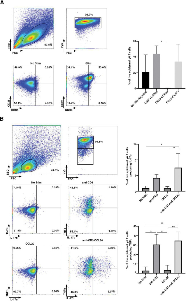
Obesity is associated with comorbidities including type 2 diabetes, chronic nonhealing wounds and psoriasis. Normally skin homeostasis and repair is regulated through the production of cytokines and growth factors derived from skin-resident cells including epidermal γδ T cells. However epidermal γδ T cells exhibit reduced proliferation and defective growth factor and cytokine production during obesity and type 2 diabetes. One of the genes modulated in epidermal γδ T cells during obesity and type 2 diabetes is CCR6, which is the receptor for CCL20. CCL20 is elevated in the skin during obesity and type 2 diabetes. Here we identify a subset of murine epidermal γδ T cells that expresses CCR6 in response to activation in vitro and post-wounding or psoriasis induction with imiquimod in vivo. We show that CCL20 stimulates epidermal γδ T cells to produce IL-17 suggesting CCR6 regulates the IL-17 axis as in dermal γδ T cells. Further, epidermal γδ T cells upregulate CCR6 and produce IL-17 during murine models of wound repair and psoriasis. Obesity increases CCR6 and IL-17 expression by epidermal γδ T cells during wound repair but has less of an effect during psoriasis. These findings have novel implications for the regulation of a specific population of IL-17-producing epidermal γδ T cells during skin damage and inflammation.


Figures

Figure 1.
Figure 1.. CCR6 is upregulated by a subset of CD25+ epidermal γδ T cells upon anti-CD3 stimulation.
(A) Flow cytometric analysis of epidermal cells isolated from B6 mice, cultured for 9–15 weeks (> 95% epidermal γδ T cells), and stimulated with anti-CD3. Live, γδ TCR+ cells are gated and CCR6 and CD25 analyzed (n=3). (B) Flow cytometric analysis of epidermal Vγ5 T cells stimulated in the presence or absence of anti-CD3 and/or CCL20 for 6 hours. Live, Vγ5+ T cells are gated and TNF-α and IL-17A analyzed (n=3). Data represents the mean +/− SD. *p < .05 and ** p < .01
Figure 2.
Figure 2.. CCR6+ epidermal γδ T cells exhibit a Tγδ17 gene expression profile.
Using publicly available scRNA-Seq data (20) the total skin cell population (n=18,040) was filtered to epidermal γδ T cells (n=236). (A) UMAP plot showing epidermal γδ T cells can be identified in three distinct clusters. Based on treatment group and CCR6 expression epidermal γδ T cells were clustered further into CCR6+IMQ, CCR6+control, CCR6IMQ, CCR6control. In the treatment group, the ratio of CCR6+ to CCR6 cells increased from 1/48 in the control group to 1/9. (B) Differential gene expression between 4 different epidermal γδ T cell populations within treatment groups displayed in a heatmap (C) Dual heatmap rendering of differential gene expression between specific epidermal γδ T cell groups.
Figure 3.
Figure 3.. Canonical pathway analysis of differentially expressed genes from CCR6+ and CCR6 epidermal γδ T cells during IMQ-induced psoriasis identify Myc pathway.
(A) Top differentially expressed genes between CCR6+IMQ and CCR6IMQ from publicly available scRNA sequencing data (20) were clustered into canonical pathways using IPA Knowledge Base platform. Positive z-score (orange) shows pathway activation. A negative z-score (blue) shows the pathway is inhibited. No z-score (white) shows the pathway is neither activated nor inhibited. Bars are arranged by statistical significance. (B) Network analysis identifies upstream regulator Myc (orange indicating activation), with IPA Knowledge Base predicting a linked activation with Rora. (C) Network downstream analysis of Rora pathway with orange predicting activation, blue predicting inhibition, and red predicting an increased measurement between subsets (CCR6+IMQ vs. CCR6IMQ epidermal γδ T cells).
Figure 4.
Figure 4.. IMQ-induced psoriasis increases IL-17A and CCR6 expression by epidermal γδ T cells, while obesity does not further increase expression.
(A) Representative immunofluorescent images of epidermal sheets from C57BL/6-Il17atm1Bcgen/J mice treated with and without IMQ for two days. Scale bars, 100 μm. (B) Quantification of IL-17A+, CCR6+, and IL-17A+CCR6+ epidermal γδ T cells with and without IMQ treatment (n=3 mice/group). A minimum of seven fields of view for each mouse were used for analysis and then averaged to form one data point. * p < 0.05
Figure 5.
Figure 5.. CCR6 is upregulated within the first 24 hrs. post-wounding and downregulated by 72 hrs. post-wounding.
(A) Representative immunofluorescent images of epidermal sheets from B6 mice at various timepoints post wounding. Wound site indicated with dotted line. Scale bars,100 μm. (B) Quantification of CCR6+ epidermal γδ T cells at different timepoints post wounding (n= 3–4 mice/group). A minimum of seven fields of view for each mouse were used for analysis and then averaged to form one data point. p = .05
Figure 6.
Figure 6.. IL-17A and CCR6 expression by epidermal γδ T cells is significantly elevated in wounded mice, while obesity increases IL-17A production and CCR6 expression by epidermal γδ T cells at the wound site.
(A) Representative immunofluorescent images of epidermal sheets from C57BL/6-Il17atm1Bcgen/J mice one day post wounding. Wound site indicated with dotted line. Scale bars, 100 μm. (B) Quantification of IL-17A+, CCR6+, and IL-17A+CCR6+ epidermal γδ T cells with and without wounding (n=3 mice/group). A minimum of seven fields of view for each mouse were used for analysis and then averaged to form one data point. * p < 0.05, ** p < .01, *** p < .001

Similar articles

References

    1. Abramczyk R., Queller J. N., Rachfal A. W., and Schwartz S. S.. 2020. Diabetes and Psoriasis: Different Sides of the Same Prism. Diabetes Metab. Syndr. Obes. Targets Ther. 13: 3571–3577. - PMC - PubMed
    1. Brazzelli V., Maffioli P., Bolcato V., Ciolfi C., D’Angelo A., Tinelli C., and Derosa G.. 2021. Psoriasis and Diabetes, a Dangerous Association: Evaluation of Insulin Resistance, Lipid Abnormalities, and Cardiovascular Risk Biomarkers. Front. Med. 8: 605691. - PMC - PubMed
    1. Loots M. A., Lamme E. N., Mekkes J. R., Bos J. D., and Middelkoop E.. 1999. Cultured fibroblasts from chronic diabetic wounds on the lower extremity (non-insulin-dependent diabetes mellitus) show disturbed proliferation. Arch. Dermatol. Res. 291: 93–99 - PubMed
    1. Evans E. A., Sayers S. R., Kodji X., Xia Y., Shaikh M., Rizvi A., Frame J., Brain S. D., Philpott M. P., Hannen R. F., and Caton P. W.. 2020. Psoriatic skin inflammation induces a pre-diabetic phenotype via the endocrine actions of skin secretome. Mol. Metab. 41: 101047. - PMC - PubMed
    1. Sen C. K. 2021. Human Wound and Its Burden: Updated 2020 Compendium of Estimates. Adv. Wound Care. 10: 281–292 - PMC - PubMed

Publication types