[Preprint]. 2024 Apr 5:rs.3.rs-4210090.
doi: 10.21203/rs.3.rs-4210090/v1.

Respiratory viral infection promotes the awakening and outgrowth of dormant metastatic breast cancer cells in lungs

Affiliations

Respiratory viral infection promotes the awakening and outgrowth of dormant metastatic breast cancer cells in lungs

Shi B Chia et al. Res Sq. .

Update in

  • Respiratory viral infections awaken metastatic breast cancer cells in lungs.
    Chia SB, Johnson BJ, Hu J, Valença-Pereira F, Chadeau-Hyam M, Guntoro F, Montgomery H, Boorgula MP, Sreekanth V, Goodspeed A, Davenport B, De Dominici M, Zaberezhnyy V, Schleicher WE, Gao D, Cadar AN, Petriz-Otaño L, Papanicolaou M, Beheshti A, Baylin SB, Guarnieri JW, Wallace DC, Costello JC, Bartley JM, Morrison TE, Vermeulen R, Aguirre-Ghiso JA, Rincon M, DeGregori J. Chia SB, et al. Nature. 2025 Sep;645(8080):496-506. doi: 10.1038/s41586-025-09332-0. Epub 2025 Jul 30. Nature. 2025. PMID: 40739350 Free PMC article.

Abstract

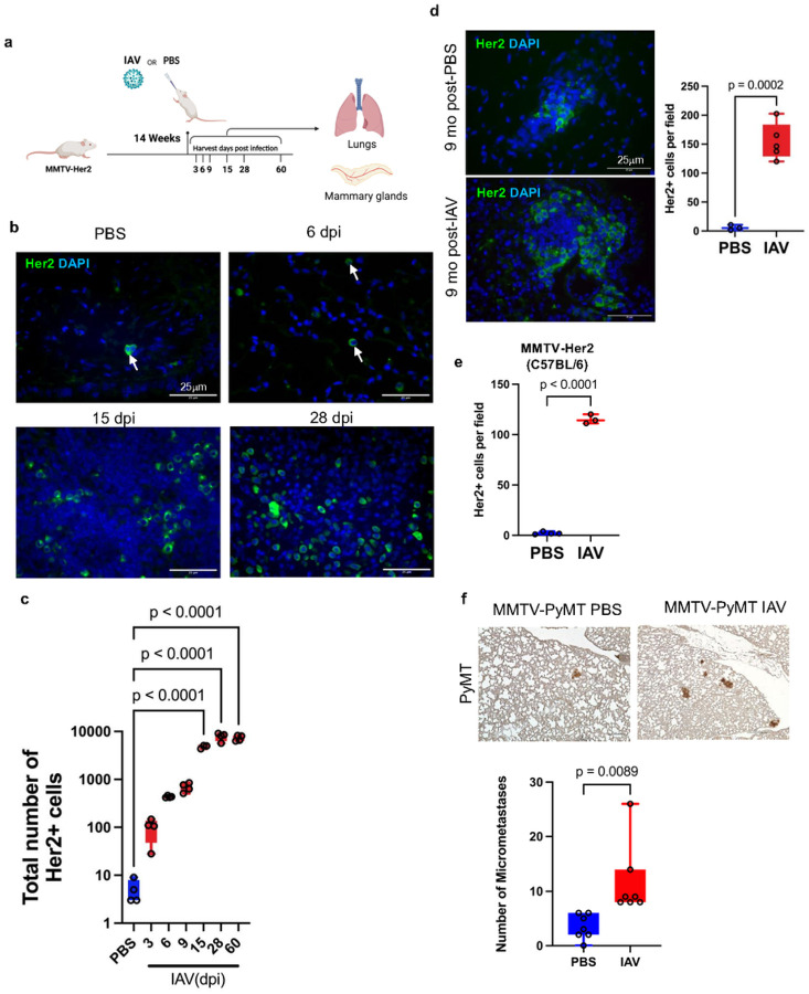
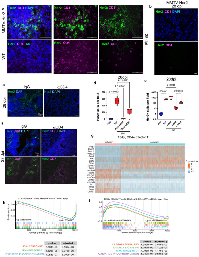
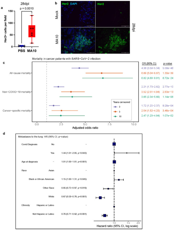
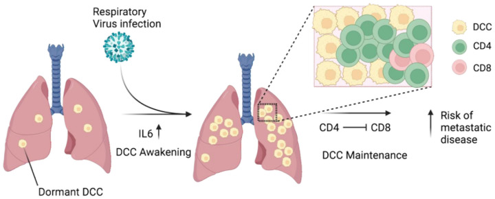
Breast cancer is the second most common cancer globally. Most deaths from breast cancer are due to metastatic disease which often follows long periods of clinical dormancy1. Understanding the mechanisms that disrupt the quiescence of dormant disseminated cancer cells (DCC) is crucial for addressing metastatic progression. Infection with respiratory viruses (e.g. influenza or SARS-CoV-2) is common and triggers an inflammatory response locally and systemically2,3. Here we show that influenza virus infection leads to loss of the pro-dormancy mesenchymal phenotype in breast DCC in the lung, causing DCC proliferation within days of infection, and a greater than 100-fold expansion of carcinoma cells into metastatic lesions within two weeks. Such DCC phenotypic change and expansion is interleukin-6 (IL-6)-dependent. We further show that CD4 T cells are required for the maintenance of pulmonary metastatic burden post-influenza virus infection, in part through attenuation of CD8 cell responses in the lungs. Single-cell RNA-seq analyses reveal DCC-dependent impairment of T-cell activation in the lungs of infected mice. SARS-CoV-2 infected mice also showed increased breast DCC expansion in lungs post-infection. Expanding our findings to human observational data, we observed that cancer survivors contracting a SARS-CoV-2 infection have substantially increased risks of lung metastatic progression and cancer-related death compared to cancer survivors who did not. These discoveries underscore the significant impact of respiratory viral infections on the resurgence of metastatic cancer, offering novel insights into the interconnection between infectious diseases and cancer metastasis.


Conflict of interest statement

Additional Declarations: Yes there is potential Competing Interest. HM has consulted Astra Zeneca relating to the use of monoclonal antibodies in the prevention and treatment of SARS-CoV-2 infection. J.A.A-G is a scientific co-founder, scientific advisory board member and equity owner in HiberCell and receives financial compensation as a consultant for HiberCell, a Mount Sinai spin-off company focused on the research and development of therapeutics that prevent or delay the recurrence of cancer. J.A.A-G is also a consultant for Astrin Biosciences, Inc and chief mission officer of Samuel Waxman Cancer Research Foundation.

Figures

Figure 1
Figure 1. Influenza A virus infection increases DCC in lungs.
MMTV-Her2 female mice (N=3–5/group) in FVB background were infected with a sublethal dose of Puerto Rico A/PR/8/34 H1N1 influenza A virus (IAV) by intranasal administration. Lungs and mammary glands were harvested at timepoints indicated post-infection (a) Immunofluorescence and quantification of Her2+ cells in lungs 3, 6, 9, 15, 28, 60 dpi, with total numbers from three sections of the whole lung quantified. (b, c) Lung sections were stained with DAPI (blue), and Her2 (green) as a marker for DCC (b). Immunofluorescence and quantification of Her2+ cells in lungs 9 months post-influenza infection (d). Quantification of Her2+ cells in C57BL6/J MMTV-Her2 mouse lungs at 15dpi with IAV (N=4 PBS, 3 IAV) (e). Immunohistochemistry and quantification of PyMT+ micrometastases defined by lesions with an area < 0.03 mm2 (N=7/group) (f). Significance determined by one-way ANOVA test.
Figure 2
Figure 2. Influenza A virus infection promotes dormant DCC proliferation and induces phenotypic changes through IL-6.
Immunofluorescence and quantification of Ki67+/Her2+ cells in lungs post-influenza A virus (IAV) infection. Lung sections from naïve and IAV-infected mice were stained with antibodies against Her2 (green) and Ki67 (magenta), and DAPI (blue) (a): percentage of Ki67+/Her2+ cells (b, left) and absolute number of Ki67+/Her2+ cells across three lung sections (b, right). Immunofluorescence and quantification of vimentin+ (Vim+) and EpCAM+Her2+ cells in lungs post-influenza infections where lung sections from naïve and IAV-infected mice were stained with Her2 (green) and vimentin (magenta) (c) or Her2 (green) and EpCAM (magenta) (e), and percentage of vimentin+/Her2+ or EpCAM+/Her2+ cells graphed (d, f). Lung sections of MMTV-Her2 or IL-6 KO:MMTV-Her2 mice 28dpi (PBS or IAV) were stained for Her2 (green) and DAPI (blue) (g) and quantified at 9- and 28dpi (h, i). Quantification of the percentage of Ki67+Her2+ cells in MMTV-Her2 and IL-6 KO:MMTV-Her2 9dpi (j). Quantification of the percentage of vimentin+/Her2+ cells in MMTV-Her2 and IL-6 KO:MMTV-Her2 mice at 9- and 28dpi (k). 3–5 mice were used in each group. Statistical Significance relative to PBS samples are shown, as determined by one-way ANOVA test.
Figure 3
Figure 3. CD4+ cells are required to maintain IAV-mediated Her2+ DCC expansion.
Adjacent lung sections of IAV-infected mice 28dpi were stained for Her2 (green) and CD4 (red, middle) or Her2 (green) and CD8 (red, right) (a). As in (a) for IAV-infected MMTV-Her2 mice 28 dpi, but for a region devoid of CD4 cells (b). Lung sections of MMTV-Her2 or CD4-depleted-MMTV-Her2 mice 28 dpi were stained for Her2 (green) and DAPI (blue) (c). Number of Her2+ cells were quantified from IAV-infected mice with CD4 depleted −1 and 10dpi (d). Quantification of Her2+ cells from MMTV-Her2 mice with CD4, CD8, CD4/8 depletion (on −1dpi) at 28dpi (e). Lung sections of MMTV-Her2 and CD4-depleted MMTV-Her2 mice at 28dpi were stained for Her2 (green), CD8 (magenta), and DAPI (blue) (f). 3–5 mice were used in each group. p determined by one-way ANOVA test. Heatmap of top 20 differentially expressed genes from scRNAseq comparing CD4 effector T cells from MMTV-Her2+IAV versus WT+IAV mice at 15 dpi (g). GSEA analyses comparing effector CD4 T cells in MMTV-Her2+IAV versus WT+IAV mice at 15 dpi (h), and effector CD8+ T cells in MMTV-Her2+IAV versus CD4-depleted MMTV-Her2+IAV mice at 15 dpi (i).
Figure 4
Figure 4. SARS-CoV-2 infection increases cancer progression and metastasis to lungs.
Quantification of Her2+ cells (a) across three lung sections in C57BL6/J MMTV-Her2 mouse lungs at 28dpi with MA10 SARS-CoV-2 (N=6) or vehicle (PBS; N=7). Immunofluorescent stain of Her2 at 28dpi (b). Analyses of UK Biobank for the association of cancer patients tested positive for SARS-CoV-2 or not and the risk of cancer-related mortality (c). Analyses of the Flatiron Health database with the hazard ratio of risks of progression to metastatic disease in the lungs between breast cancer patients tested COVID-positive or not (d).
Figure 5
Figure 5. Model.
Pulmonary virus-dependent increases in IL-6 contribute to the awakening and expansion of dormant mesenchymal-like breast cancer cells that switch to a partial epithelial-like phenotype in lungs in the early phase of viral infection. CD4 cells maintain the expanded breast cancer cells in late phase of viral infection through suppressing CD8 cells. Virus-dependent awakening and expansion of DCC in the lungs increases the risks of metastatic progression.

References

    1. Siegel R. L., Miller K. D. & Jemal A. Cancer statistics, 2020. CA Cancer J Clin 70, 7–30 (2020). 10.3322/caac.21590 - DOI - PubMed
    1. Newton A. H., Cardani A. & Braciale T. J. The host immune response in respiratory virus infection: balancing virus clearance and immunopathology. Semin Immunopathol 38, 471–482 (2016). 10.1007/s00281-016-0558-0 - DOI - PMC - PubMed
    1. Tan L. Y., Komarasamy T. V. & Rmt Balasubramaniam V. Hyperinflammatory Immune Response and COVID-19: A Double Edged Sword. Front Immunol 12, 742941 (2021). 10.3389/fimmu.2021.742941 - DOI - PMC - PubMed
    1. Soni A. et al. Breast cancer subtypes predispose the site of distant metastases. Am J Clin Pathol 143, 471–478 (2015). 10.1309/AJCPYO5FSV3UPEXS - DOI - PubMed
    1. Phan T. G. & Croucher P. I. The dormant cancer cell life cycle. Nat Rev Cancer 20, 398–411 (2020). 10.1038/s41568-020-0263-0 - DOI - PubMed

Publication types