Review
. 2024 Apr 5:11:1359426.
doi: 10.3389/fvets.2024.1359426. eCollection 2024.

Melanoma of the dog and cat: consensus and guidelines

Affiliations
Review

Melanoma of the dog and cat: consensus and guidelines

Gerry Polton et al. Front Vet Sci. .

Erratum in

  • Corrigendum: Melanoma of the dog and cat: consensus and guidelines.
    Polton G, Borrego JF, Clemente-Vicario F, Clifford CA, Jagielski D, Kessler M, Kobayashi T, Lanore D, Queiroga FL, Rowe AT, Vajdovich P, Bergman PJ. Polton G, et al. Front Vet Sci. 2024 Jul 2;11:1442751. doi: 10.3389/fvets.2024.1442751. eCollection 2024. Front Vet Sci. 2024. PMID: 39015108 Free PMC article.

Abstract

Melanoma of the dog and cat poses a clinical challenge to veterinary practitioners across the globe. As knowledge evolves, so too do clinical practices. However, there remain uncertainties and controversies. There is value for the veterinary community at large in the generation of a contemporary wide-ranging guideline document. The aim of this project was therefore to assimilate the available published knowledge into a single accessible referenced resource and to provide expert clinical guidance to support professional colleagues as they navigate current melanoma challenges and controversies. Melanocytic tumors are common in dogs but rare in cats. The history and clinical signs relate to the anatomic site of the melanoma. Oral and subungual malignant melanomas are the most common malignant types in dogs. While many melanocytic tumors are heavily pigmented, making diagnosis relatively straightforward, melanin pigmentation is variable. A validated clinical stage scheme has been defined for canine oral melanoma. For all other locations and for feline melanoma, TNM-based staging applies. Certain histological characteristics have been shown to bear prognostic significance and can thus prove instructive in clinical decision making. Surgical resection using wide margins is currently the mainstay of therapy for the local control of melanomas, regardless of primary location. Radiotherapy forms an integral part of the management of canine oral melanomas, both as a primary and an adjuvant therapy. Adjuvant immunotherapy or chemotherapy is offered to patients at high risk of developing distant metastasis. Location is the major prognostic factor, although it is not completely predictive of local invasiveness and metastatic potential. There are no specific guidelines regarding referral considerations for dogs with melanoma, as this is likely based on a multitude of factors. The ultimate goal is to provide the best options for patients to extend quality of life and survival, either within the primary care or referral hospital setting.

Keywords: cat; consensus; dog; guidelines; melanoma; prognosis; treatment.


Conflict of interest statement

The authors declare that the research was conducted in the absence of any commercial or financial relationships that could be construed as a potential conflict of interest.

Figures

Figure 1
Figure 1
Oral melanoma. (A–D) Heterogenous appearance of oral melanoma in a cocker spaniel. (A) Outward appearance. (B) Visualization of the intraoral aspect illustrates the importance of oral examination in forming the clinical suspicion of melanoma. (C,D) The intraoperative and excision specimen images reveal the absence of an appreciable surgical margin. This is acceptable when the goal of surgery is palliation of the consequences of the presence of a large mass or as a precursor to definitive radiotherapy. (D) Heterogenous appearance of oral melanoma: the tumor grossly exhibits melanotic and amelanotic parts. (E) Malignant melanoma cytology from mandibular gingiva. Cellular borders are usually distinct, though sometimes faint. Cytoplasm is basophilic or light basophilic and granulated; occasionally, small vacuoles or light black to deep green small cytoplasmic granules are seen. Nuclei are eccentrically located and may be irregular; they often show large, dark, prominent nucleoli. (F–I) Oral malignant melanoma with lymph node metastasis in a dachshund. (F) A small primary tumor was located medial to a left upper molar tooth. (G) The patient was presented with left submandibular lymph node enlargement. (H,I). Tumor cells are spindle-shaped and contain large amounts of melanin pigment. The melanoma cells are large and elongated, with length 4-5x and width 2-3x accompanying red blood cells. The cytoplasmic margins are indistinct, the cytoplasm is vacuolated and black/green cytoplasmic granules are apparent. The nuclei are round to oval with stippled, coarse chromatin; nucleoli are distinct and vary in size and number. (J) Spindle-shaped form of malignant melanoma cytologically resembles a sarcoma, though melanin granules are also evident in this sample. (K,L) Oral melanoma with lymph node metastasis; melanophages versus malignant melanoma. (K) A macrophage (melanophage) with intracytoplasmic melanin pigment (arrow) is seen among reactive lymphoid population. Free melanin granules are also visible. Melanophages in themselves do not indicate tumor metastasis, as they can be seen in reactive or normal lymph nodes from dogs without melanocytic tumors. (L) Intracytoplasmic melanin pigment is evident in a large epithelioid melanoma cell (arrow) among a population of reactive lymphoid cells. (E,I,J–L) Original magnification x 1,000. (H) Original magnification x 800.
Figure 2
Figure 2
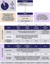
Oral melanoma decision tree.

References

    1. Gillard M, Cadieu E, De Brito C, Abadie J, Vergier B, Devauchelle P, et al. . Naturally occurring melanomas in dogs as models for non-UV pathways of human melanomas. Pigment Cell Melanoma Res. (2014) 27:90–102. doi: 10.1111/pcmr.12170, PMID: - DOI - PubMed
    1. Bergman P, Selmic LE, Kent MS. Melanoma In: Vail D, Thamm D, Liptak J, editors. Withrow and MacEwen’s small animal clinical oncology. 6th ed. St. Louis, MO: Elsevier; (2020). 367–81.
    1. Smith SH, Goldschmidt H, Mcmanus PM. A comparative review of melanocytic neoplasms. Vet Pathol. (2002) 39:651–78. doi: 10.1354/vp.39-6-651 - DOI - PubMed
    1. Goldschmidt MH. Pigmented lesions of the skin. Clin Dermatol. (1994) 12:507–14. doi: 10.1016/0738-081x(94)90217-8 - DOI - PubMed
    1. Grassinger JM, Floren A, Müller T, Cerezo-Echevarria A, Beitzinger C, Conrad D, et al. . Digital lesions in dogs: a statistical breed analysis of 2912 cases. Vet Sci. (2021) 8:136. doi: 10.3390/vetsci8070136, PMID: - DOI - PMC - PubMed