

Magnetic resonance imaging in rabies encephalitis, a case report, and review of the literature
- PMID: 38645944
- PMCID: PMC11031717
- DOI: 10.1016/j.radcr.2024.03.072
Magnetic resonance imaging in rabies encephalitis, a case report, and review of the literature
Abstract
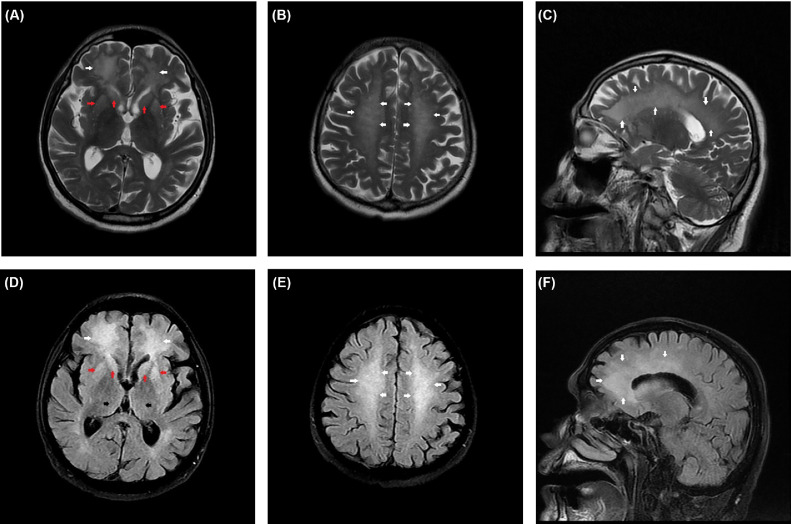
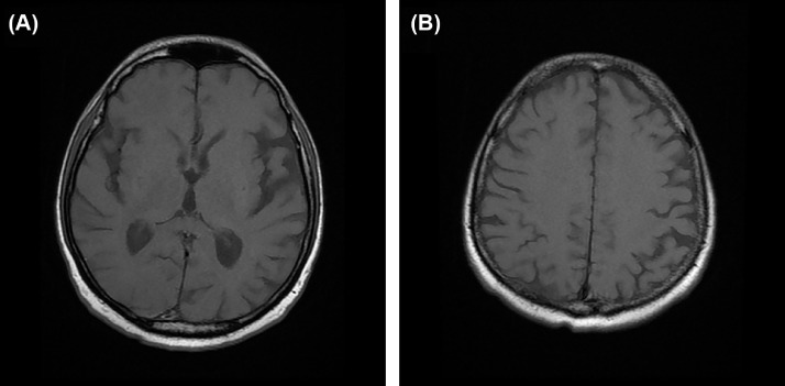
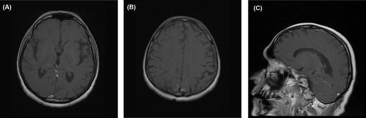
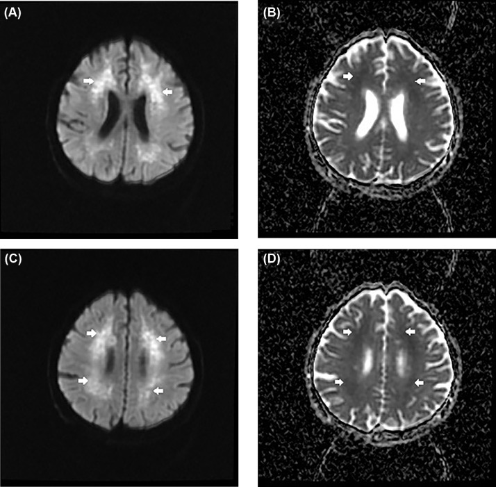
Rabies is an acute fatal disease of the central nervous system. Neuroimaging plays an important role, especially in establishing an early diagnosis and distinguishing it from other types of encephalitis. This case report aims to give a brief review of this condition and report the less common MRI findings of the disease. We herein report a case of a 61-year-old male bitten by a stray dog who presented with fever, vomiting, headache, sialorrhea, dysarthria, dysphagia, and upper limb weakness which progressed to lower limbs on the next day. T2W and FLAIR images demonstrated subtle bilateral hyperintense signal in the deep gray matter with more apparent increased signal intensity in the white matter of the frontal and parietal lobes which shows mild diffusion restriction but no postcontrast enhancement. The diagnosis of rabies encephalitis was made based on a typical history of exposure, a compatible clinical presentation, and MRI findings. Rabies diagnosis is essentially clinical. It is definitively confirmed by the isolation of the virus from biological samples such as saliva, CSF, hair, or detection of rabies antigens or antibodies. Magnetic resonance imaging (MRI) brain used as one of the modalities of investigation for distinguishing it from other encephalitis. Rabies per se does not have any characteristic features on the MRI brain.
Keywords: FLAIR; Imaging findings; MRI; Rabies encephalitis; T2WI.
© 2024 The Authors. Published by Elsevier Inc. on behalf of University of Washington.
Figures
Similar articles
-
MRI findings in human rabies: A case report on the importance of neuroimaging when biological tests are inconclusive.Radiol Case Rep. 2025 Apr 12;20(7):3281-3286. doi: 10.1016/j.radcr.2025.03.056. eCollection 2025 Jul. Radiol Case Rep. 2025. PMID: 40292140 Free PMC article.
-
Neuroimaging Findings in Rabies Encephalitis.J Neuroimaging. 2021 May;31(3):609-614. doi: 10.1111/jon.12833. Epub 2021 Feb 3. J Neuroimaging. 2021. PMID: 33534937
-
The Spectrum of MRI Findings in Dengue Encephalitis.Cureus. 2022 Sep 11;14(9):e29048. doi: 10.7759/cureus.29048. eCollection 2022 Sep. Cureus. 2022. PMID: 36237802 Free PMC article.
-
FLAIR-hyperintense lesions in anti-MOG-associated encephalitis with seizures overlaying anti-N-methyl-D-aspartate receptor encephalitis: a case report and literature review.Front Immunol. 2023 Apr 17;14:1149987. doi: 10.3389/fimmu.2023.1149987. eCollection 2023. Front Immunol. 2023. PMID: 37138864 Free PMC article.
-
Update on neuroimaging in infectious central nervous system disease.Curr Opin Neurol. 2004 Aug;17(4):475-80. doi: 10.1097/01.wco.0000137540.29857.bf. Curr Opin Neurol. 2004. PMID: 15247545 Review.
References
-
- Jameson JL, Fauci AS, Kasper DL, Hauser SL, Longo DL, Loscalzo J. 20th ed. McGraw-Hill Education; New York: 2018. Harrison’s principles of internal medicine. eds.
Publication types
LinkOut - more resources
Full Text Sources
