. 2024 Apr 10;25(1):2338785.
doi: 10.1080/14686996.2024.2338785. eCollection 2024.

Fine tuning of the net charge alternation of polyzwitterion surfaced lipid nanoparticles to enhance cellular uptake and membrane fusion potential

Affiliations

Fine tuning of the net charge alternation of polyzwitterion surfaced lipid nanoparticles to enhance cellular uptake and membrane fusion potential

Keitaro Homma et al. Sci Technol Adv Mater. .

Abstract

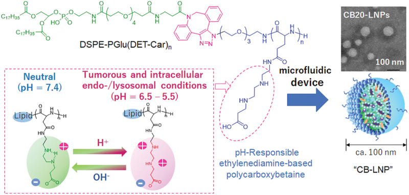
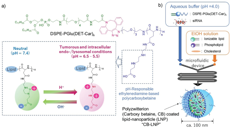
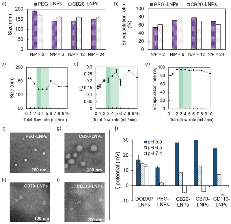
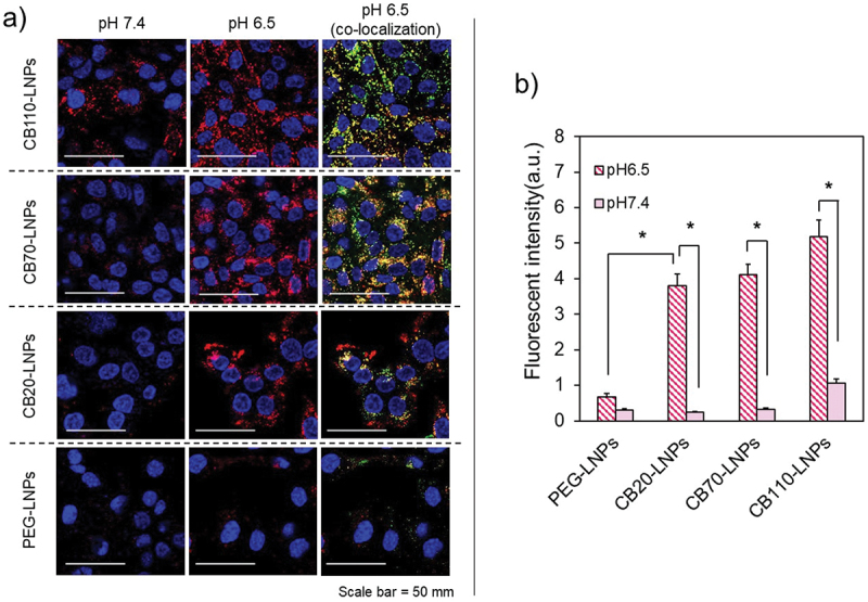
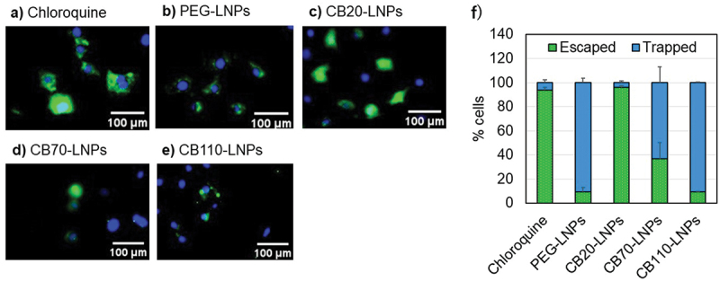
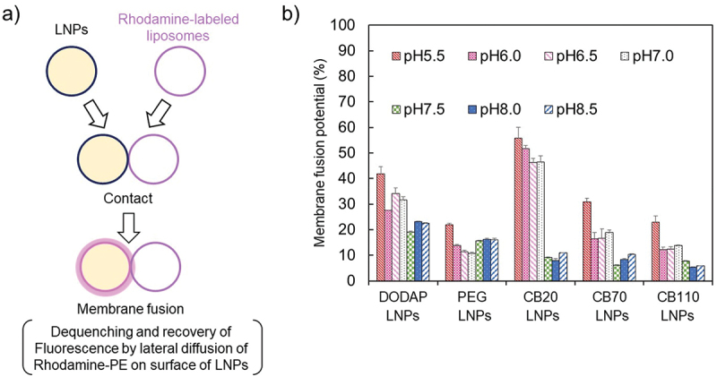
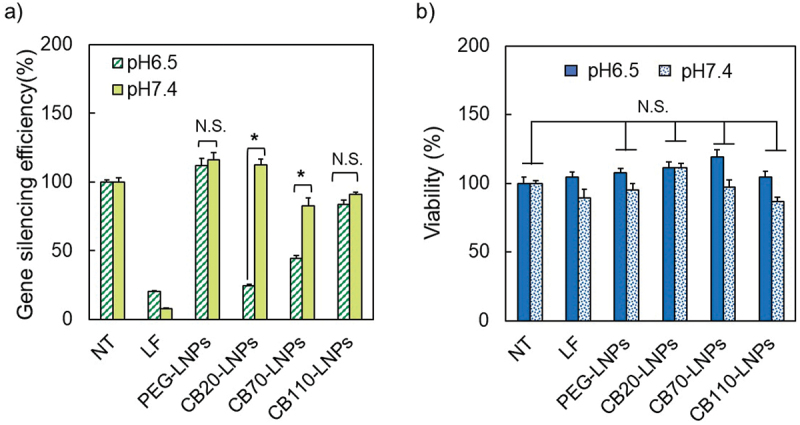
Lipid nanoparticles (LNPs) coated with functional and biocompatible polymers have been widely used as carriers to deliver oligonucleotide and messenger RNA therapeutics to treat diseases. Poly(ethylene glycol) (PEG) is a representative material used for the surface coating, but the PEG surface-coated LNPs often have reduced cellular uptake efficiency and pharmacological activity. Here, we demonstrate the effect of pH-responsive ethylenediamine-based polycarboxybetaines with different molecular weights as an alternative structural component to PEG for the coating of LNPs. We found that appropriate tuning of the molecular weight around polycarboxybetaine-modified LNP, which incorporated small interfering RNA, could enhance the cellular uptake and membrane fusion potential in cancerous pH condition, thereby facilitating the gene silencing effect. This study demonstrates the importance of the design and molecular length of polymers on the LNP surface to provide effective drug delivery to cancer cells.

Keywords: Lipid nanoparticles; membrane fusion; pH-responsiveness; polycarboxybetaine; siRNA.

Plain language summary

The study presents the unique characteristics of small interfering RNA (siRNA)-loaded lipid nanoparticles (LNPs) with different lengths of PGlu(DET-Car), revealing the length of PGlu(DET-Car) critically affects the formation of a stable LNP, the cellular uptake, membrane fusion, and gene silencing abilities.


Conflict of interest statement

No potential conflict of interest was reported by the author(s).

Figures

None
Graphical abstract
Figure 1.
Figure 1.
Preparation method of polyzwitterion coated lipid-nanoparticle (LNP) (CB-LNP). (a) Chemical structure and the protonation functionality. (b) Summary of LNP preparations using a microfluidic device.
Figure 2.
Figure 2.
Physicochemical characteristics of lipid nanoparticles (LNPs). (a,b) Initial LNPs formulated at different N/P ratios were prepared by microfluidic device with 0.5 mL/min total flow rate. (a) Size, (b) encapsulation ratio of siRNA. Data are expressed as mean ± standard deviation (SD) (n = 3). (c,e) Effect of total flow rate for preparation of LNPs. (c) Size, (d) polydispersity index (PDI), (e) encapsulation rate. (f,i) Transmission electron microscopy images. (j) Relationship between pH and ζ potential of LNPs (n = 3).
Figure 3.
Figure 3.
Confocal laser scanning microscopy (CLSM) observation of pH-dependent cellular uptake. (a) SK-OV-3-luc cells were incubated with Alexa Flour 647-siRNA conjugated lipid nanoparticles (LNPs) (red) for 72 h, and then stained for early endosome (green) and nucleus (blue). Colocalization is shown in yellow. Scale bar = 50 µm. (b) Quantitative analysis of cellular uptake. Statistical significance was evaluated using Student’s t-test, *p < 0.05. The results are expressed as means ± standard deviation (n = 30).
Figure 4.
Figure 4.
Endosomal escape analysis of LNPs in SK-OV-3-luc cells cultured at pH 6.5. (a-e) Cytosolic distribution of calcein after 2 h co-incubation of each LNPs and calcein. Cytosolic localization of calcein (green). Nuclei were stained using Hoechst 33,342 (blue). Endosomal escape is indicated by diffuse fluorescence throughout the cell, whereas punctate fluorescence represents endosomal trapping. (f) Percentage of cells showing endosomal escape (diffused fluorescence) and endosomal trapping (punctate fluorescence) as quantified using BZ-X800 Analyzer (ver. 1.1.24) (n = 30 cells). Scale bar (White) = 100 µm. The results are shown as mean ± SD.
Figure 5.
Figure 5.
Membrane fusion assay. (a) Schematic of membrane fusion assay. (b) pH-dependent potential of membrane fusion (n = 3).
Figure 6.
Figure 6.
Effect of pH on (a) gene silencing and (b) cell viability with PGlu(DET-Car)-coated lipid nanoparticles (LNPs) against SK-OV-3-luc cells. NT: Non-treatment, LF: Lipofectamine RNAi MAX. (n = 3–6). Statistical significance was evaluated using Student’s t-test, *p < 0.05. The results are expressed as means ± standard deviation.

Similar articles

Cited by

References

    1. Kulkarni JA, Witzigmann D, Thomson SB, et al. The current landscape of nucleic acid therapeutics. Nat Nanotech. 2021;16(6):630–12. doi: 10.1038/s41565-021-00898-0 - DOI - PubMed
    1. Hu B, Weng Y, Xia XH, et al. Clinical advances of siRNA therapeutics. J Gene Med. 2019;21(7):e3097. doi: 10.1002/jgm.3097 - DOI - PubMed
    1. Mitchell MJ, Billingsley MM, Haley RM, et al. Engineering precision nanoparticles for drug delivery. Nat Rev Drug Discov. 2020;20(2):101–124. - PMC - PubMed
    1. Urits I, Swanson D, Swett MC, et al. A review of Patisiran (ONPATTRO®) for the treatment of polyneuropathy in people with hereditary transthyretin amyloidosis. Neurol Ther. 2020;9(2):301–315. - PMC - PubMed
    1. Wang X. Safety and efficacy of the BNT162b2 mRNA Covid-19 vaccine. N Engl J Med. 2021;383:2603–2615. doi: 10.1056/NEJMc2036242 - DOI - PMC - PubMed

LinkOut - more resources