

Chronic Visual Abnormality in an Elderly Patient With Temporal Lobe Epilepsy
- PMID: 38646321
- PMCID: PMC11032511
- DOI: 10.7759/cureus.56696
Chronic Visual Abnormality in an Elderly Patient With Temporal Lobe Epilepsy
Abstract
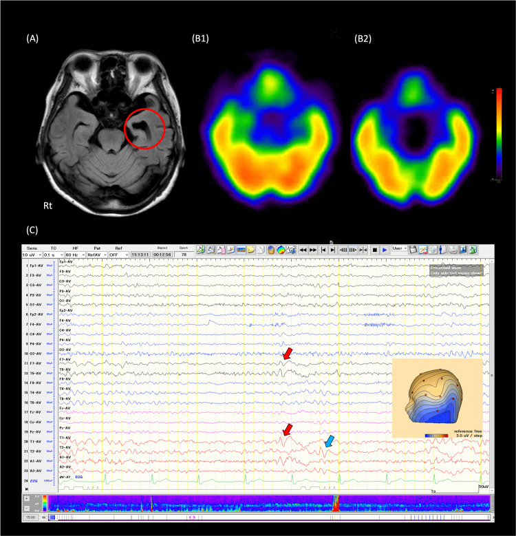
A 79-year-old woman visited our department for chronic visual field abnormalities with a floating sensation for two months. Neurological and ophthalmologic examinations yielded normal results, except for brain MRI indicating left hippocampal atrophy. Cognitive function tests were normal. EEG revealed frequent spikes and slow waves in the left frontotemporal region, corroborated by reduced accumulation in 123I-iomazenil single photon emission computed tomography. A diagnosis of temporal lobe epilepsy was established, and treatment with lacosamide resulted in a remarkable improvement in symptoms and EEG findings. Mild focal seizures from the temporal region might cause mild impaired awareness, resulting in the patient's report as a sensation of uncertainty between the self and the outside world, mimicking ophthalmologic abnormalities. The repeated nature of the seizures contributed to the absence of the term "transient" in symptom description. Diagnosing epilepsy in the elderly proves challenging due to nonspecific complaints.
Keywords: clinical neurophysiology; elderly; electroencephalography; focal seizure; loss of awareness.
Copyright © 2024, Atsuji et al.
Conflict of interest statement
The authors have declared that no competing interests exist.
Figures
Similar articles
-
An analysis of clinical seizure patterns and their localizing value in frontal and temporal lobe epilepsies.Brain. 1996 Feb;119 ( Pt 1):17-40. doi: 10.1093/brain/119.1.17. Brain. 1996. PMID: 8624679
-
Is 11C-flumazenil PET superior to 18FDG PET and 123I-iomazenil SPECT in presurgical evaluation of temporal lobe epilepsy?J Neurol Neurosurg Psychiatry. 1997 Feb;62(2):141-50. doi: 10.1136/jnnp.62.2.141. J Neurol Neurosurg Psychiatry. 1997. PMID: 9048714 Free PMC article.
-
Improvement of epilepsy with lacosamide in a patient with ring chromosome 20 syndrome.Brain Dev. 2020 Jun;42(6):473-476. doi: 10.1016/j.braindev.2020.03.003. Epub 2020 Apr 1. Brain Dev. 2020. PMID: 32247529
-
Current management and surgical outcomes of medically intractable epilepsy.Clin Neurol Neurosurg. 2013 Dec;115(12):2411-8. doi: 10.1016/j.clineuro.2013.09.035. Epub 2013 Oct 11. Clin Neurol Neurosurg. 2013. PMID: 24169149 Review.
-
The usefulness of stereo-electroencephalography (SEEG) in the surgical management of focal epilepsy associated with "hidden" temporal pole encephalocele: a case report and literature review.Neurosurg Rev. 2018 Jan;41(1):347-354. doi: 10.1007/s10143-017-0922-0. Epub 2017 Oct 16. Neurosurg Rev. 2018. PMID: 29039074 Review.
References
-
- Detection of hippocampal atrophy in patients with temporal lobe epilepsy: a 3-Tesla MRI shape. Mumoli L, Labate A, Vasta R, et al. Epilepsy Behav. 2013;28:489–493. - PubMed
Publication types
LinkOut - more resources
Full Text Sources