

When Tuberculosis Defies Appearances: The Tale of a Deceptive Abdominal Mass on Imaging
- PMID: 38646345
- PMCID: PMC11032693
- DOI: 10.7759/cureus.56686
When Tuberculosis Defies Appearances: The Tale of a Deceptive Abdominal Mass on Imaging
Abstract
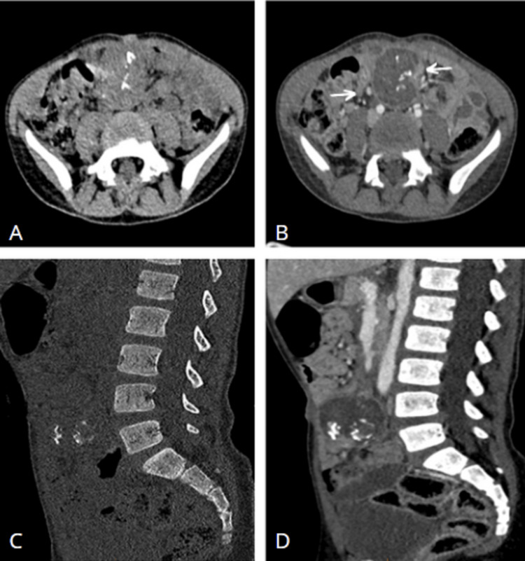
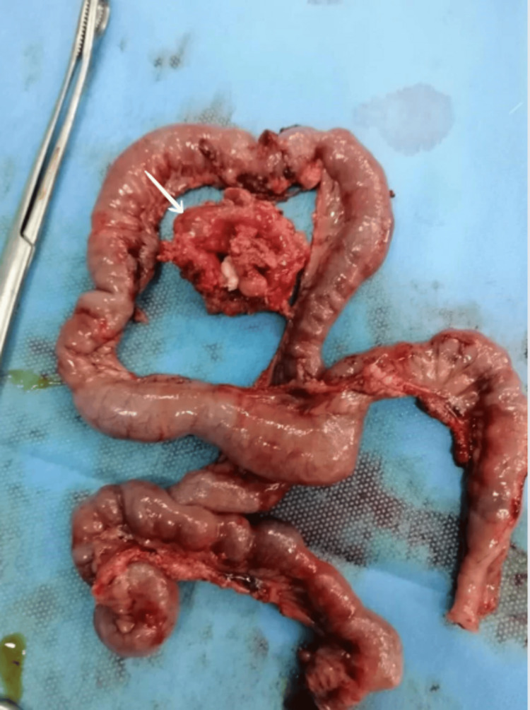
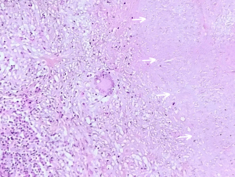
Tuberculosis poses a significant public health challenge, especially in highly endemic countries. Rarely, it appears as an abdominal mass resembling a malignant abdominal tumor and can be misleading on imaging, so early diagnosis remains a challenge, and confirmation may require invasive examinations such as laparotomy. The most characteristic radiological appearance is that of a solid, hypervascular, or peripherally enhancing mass with a hypodense center. We present a case of retroperitoneal tuberculosis that simulated a teratoma on imaging. This case highlights the diagnosis difficulties even in endemic countries, despite advances in imaging techniques such as ultrasound and computed tomography.
Keywords: abdomen; computed tomography; mass; pseudotumor; tuberculosis.
Copyright © 2024, Tkak et al.
Conflict of interest statement
The authors have declared that no competing interests exist.
Figures
References
-
- Abdominal tuberculosis of pseudotumor aspect [Article in French] Romand F, Gaudin JL, Bobichon R, Souquet JC. https://pubmed.ncbi.nlm.nih.gov/9452735/ Presse Med. 1997;26:1717–1721. - PubMed
-
- Pseudotumoral form of abdominal tuberculosis: report of four cases [Article in French] Hablani N, Souei Mhiri M, Tlili Graies K, Jemni Gharbi H, Abdallah S, Bel Hadj Hamida R. J Radiol. 2005;86:1021–1025. - PubMed
-
- Abdominal pseudotumoral tuberculosis [Article in French] El Barni R, Lahkim M, Achour A. https://pubmed.ncbi.nlm.nih.gov/23330023/ Pan Afr Med J. 2012;13:32. - PMC - PubMed
-
- Extrapulmonary tuberculosis in the central western region. Retrospective study of 217 cases (Gericco 1991-1993) [Article in French] Denis-Delpierre N, Merrien D, Billaud E, et al. https://pubmed.ncbi.nlm.nih.gov/9767996/ Presse Med. 1998;27:341–346. - PubMed
Publication types
LinkOut - more resources
Full Text Sources