

The clinical challenge of a uterine cotyledonoid dissecting leiomyoma with adenomyosis: A case report
- PMID: 38646502
- PMCID: PMC11031714
- DOI: 10.1016/j.crwh.2024.e00604
The clinical challenge of a uterine cotyledonoid dissecting leiomyoma with adenomyosis: A case report
Abstract
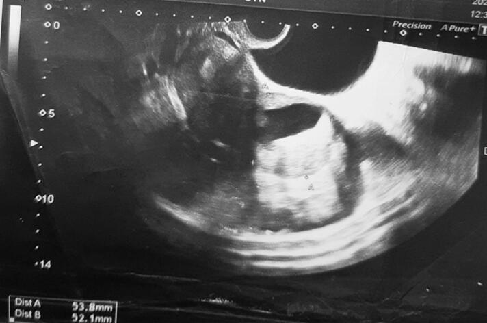
Cotyledonoid dissecting leiomyoma (CDL) is a rare uterine tumor with unique clinical and histological features. We present a case of a 46-year-old woman with a 3-month history of left-flank pain radiating to the back. The patient had a history of infertility and a previous miscarriage. Ultrasound revealed a solid tissue mass suggestive of a degenerated fibroid. Laparoscopy identified subserosal leiomyoma and leiomyoma in the broad ligament. Histologically, CDL is characterized by disorganized smooth muscle with hyaline degeneration and no evidence of malignancy. Clinically, CDL can present with a variety of symptoms, including heavy menstrual bleeding, pelvic pain, and infertility. The coexistence of CDL and adenomyosis is exceedingly rare. This case highlights the importance of considering CDL in the differential diagnosis of pelvic mass, malignant neoplasms, and infertility, even with atypical symptoms. It also emphasizes the value of cooperation between clinicians and pathologists for accurate diagnosis and management of CDL. Adenomyosis in this case further complicated the diagnosis and highlighted the need for an index of suspicion for this rare condition.
Keywords: Adenomyosis; Case report; Cotyledonoid dissecting leiomyoma; Leiomyoma; Uterine tumor.
© 2024 The Authors. Published by Elsevier B.V.
Conflict of interest statement
The authors declare that they have no conflict of interest regarding the publication of this case report.
Figures


Similar articles
-
An unusual case of uterine cotyledonoid dissecting leiomyoma with adenomyosis.Diagn Pathol. 2016 Aug 4;11(1):69. doi: 10.1186/s13000-016-0523-1. Diagn Pathol. 2016. PMID: 27491369 Free PMC article.
-
Cotyledonoid Dissecting Leiomyoma: A Rare Variant of Leiomyoma of the Uterus.Cureus. 2022 Oct 16;14(10):e30352. doi: 10.7759/cureus.30352. eCollection 2022 Oct. Cureus. 2022. PMID: 36407217 Free PMC article.
-
Case report: cotyledonoid dissecting leiomyoma in a 49-year-old woman.Transl Cancer Res. 2022 Nov;11(11):4189-4193. doi: 10.21037/tcr-22-1521. Transl Cancer Res. 2022. PMID: 36523320 Free PMC article.
-
Cotyledonoid leiomyoma: a benign uterine tumor with alarming gross appearance.Arch Pathol Lab Med. 2002 Feb;126(2):210-3. doi: 10.5858/2002-126-0210-CL. Arch Pathol Lab Med. 2002. PMID: 11825122 Review.
-
Cotyledonoid dissecting leiomyoma of the uterus: a case report and review of the literature.J Med Case Rep. 2023 Dec 16;17(1):516. doi: 10.1186/s13256-023-04271-8. J Med Case Rep. 2023. PMID: 38102631 Free PMC article. Review.
References
-
- Roth L.M., Reed R.J., Sternberg W.H. Cotyledonoid dissecting leiomyoma of the uterus. The Sternberg tumor. Am. J. Surg. Pathol. 1996 Dec;20(12):1455–1461. - PubMed
-
- Gezginç K., Yazici F., Selimoğlu R., Tavli L. Cotyledonoid dissecting leiomyoma of the uterus with intravascular growth in postmenopausal woman: a case presentation. Int. J. Clin. Oncol. 2011 Dec;16(6):701–704. - PubMed
Publication types
LinkOut - more resources
Full Text Sources
