. 2024 Apr 5:15:1382318.
doi: 10.3389/fimmu.2024.1382318. eCollection 2024.

Mucosal immunization with a low-energy electron inactivated respiratory syncytial virus vaccine protects mice without Th2 immune bias

Affiliations

Mucosal immunization with a low-energy electron inactivated respiratory syncytial virus vaccine protects mice without Th2 immune bias

Valentina Eberlein et al. Front Immunol. .

Abstract

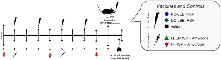
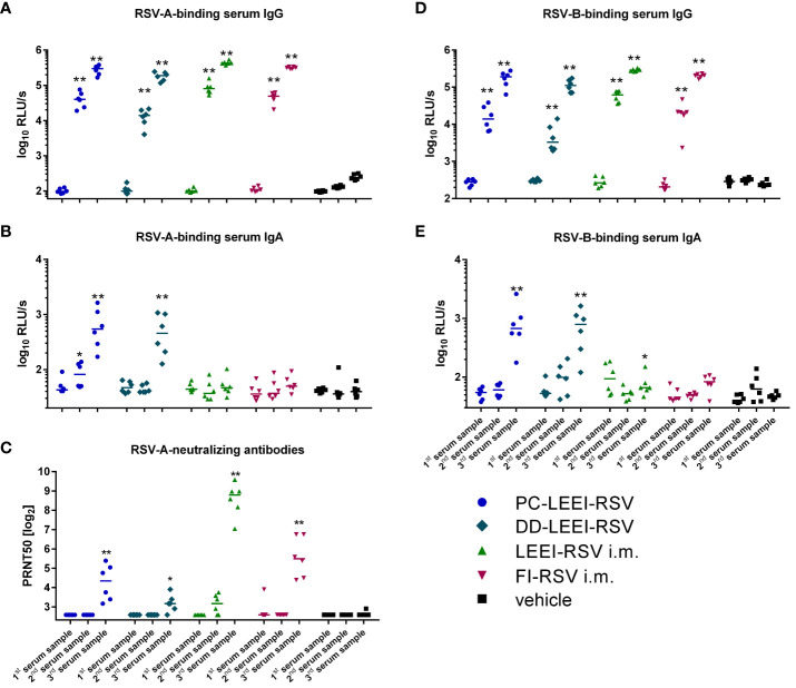
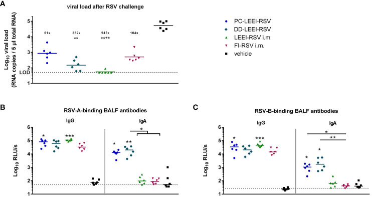
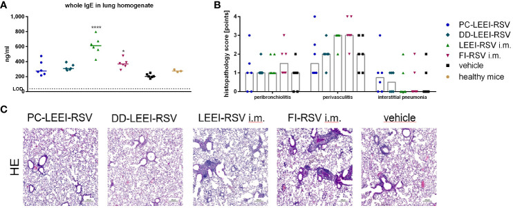
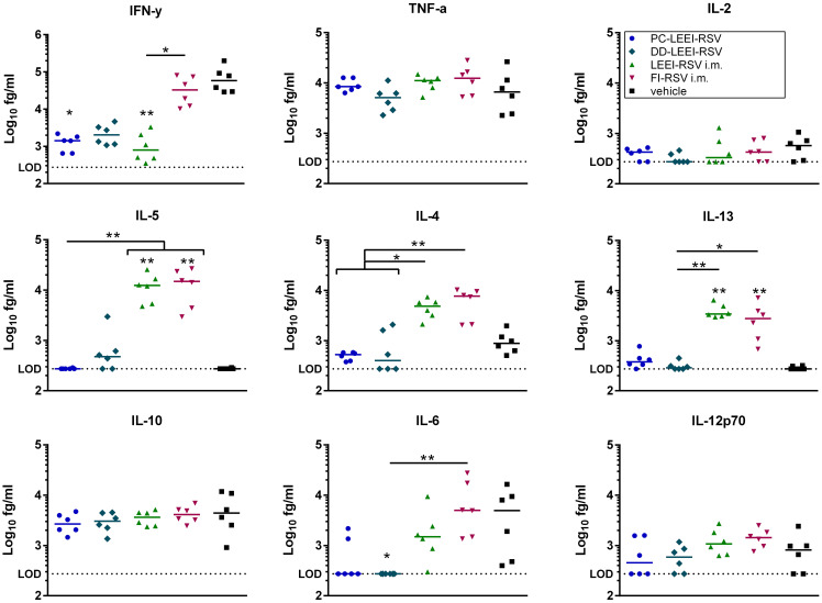
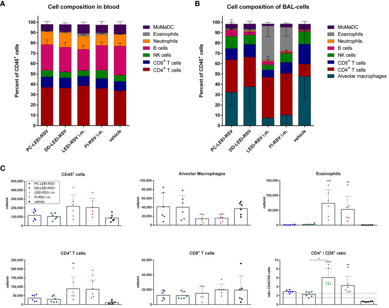
The respiratory syncytial virus (RSV) is a leading cause of acute lower respiratory tract infections associated with numerous hospitalizations. Recently, intramuscular (i.m.) vaccines against RSV have been approved for elderly and pregnant women. Noninvasive mucosal vaccination, e.g., by inhalation, offers an alternative against respiratory pathogens like RSV. Effective mucosal vaccines induce local immune responses, potentially resulting in the efficient and fast elimination of respiratory viruses after natural infection. To investigate this immune response to an RSV challenge, low-energy electron inactivated RSV (LEEI-RSV) was formulated with phosphatidylcholine-liposomes (PC-LEEI-RSV) or 1,2-dioleoyl-3-trimethylammonium-propane and 1,2-dioleoyl-sn-glycero-3-phosphoethanolamine (DD-LEEI-RSV) for vaccination of mice intranasally. As controls, LEEI-RSV and formalin-inactivated-RSV (FI-RSV) were used via i.m. vaccination. The RSV-specific immunogenicity of the different vaccines and their protective efficacy were analyzed. RSV-specific IgA antibodies and a statistically significant reduction in viral load upon challenge were detected in mucosal DD-LEEI-RSV-vaccinated animals. Alhydrogel-adjuvanted LEEI-RSV i.m. showed a Th2-bias with enhanced IgE, eosinophils, and lung histopathology comparable to FI-RSV. These effects were absent when applying the mucosal vaccines highlighting the potential of DD-LEEI-RSV as an RSV vaccine candidate and the improved performance of this mucosal vaccine candidate.

Keywords: Respiratory Syncytial Virus (RSV); formulation; low-energy electron irradiation (LEEI); mucosal immunity; mucosal vaccination.


Conflict of interest statement

SU and MT are authors of patents and patent applications covering LEEI to inactivate liquids: WO2018041953, DE102015224206B3, US10080795, DE 102017002645.9. The remaining authors declare that the research was conducted in the absence of any commercial or financial relationships that could be construed as a potential conflict of interest.

Figures

Figure 1
Figure 1
Scheme of the vaccination schedule and infection with RSV. BALB/c mice were vaccinated using homologous vaccine regiments. For the formulated LEEI-RSV vaccine with different liposomes for the formulated LEEI-RSV vaccines with different liposomes the application route was intranasal (i.n.), as well as for the vehicle group which recieved PBS with 12% trehalose. As presented, the intranasal application was performed four times in total, i.e., every 2 weeks. For comparison, the vaccinees received LEEI-RSV (LEEI-RSV i.m.) or formalin-inactivated RSV (FI-RSV i.m.) two times intramuscularly (i.m.) in a 4-week interval, both with the Alhydrogel adjuvant. Blood samples were collected for immunogenicity analysis at the indicated timepoints. At 8 weeks after the first vaccination, the animals were challenged with 106 FFU (focus-forming units) RSV and scored for 5 days to test the protective efficacy. At 5 days after challenge, the mice were euthanized and final blood samples were taken; bronchoalveolar lavage (BAL) was performed as well, whereby the left lung lobes was separated after inflation for histological analysis. The remaining lung tissue was homogenized for further analysis.
Figure 2
Figure 2
Conservation of RSV surface proteins after irradiation. The conservation of RSV-F (18F12), RSV-preF (D25), and RSV-G (8C5) after inactivation was measured in untreated control (untreated), the LEEI process control (0 mA), and after inactivation with 1.25 mA LEEI or formalin (FI) by ELISA. Shown are LEEI-RSV inactivated in MFC and formalin-inactivated RSV (FI-RSV) used in the animal-experiments in comparison to the respective controls. The bars indicate the mean of each group; every dot is a replicate, and shown is a representative experiment out of two. The calculated fold reduction to the respective 0 mA control is indicated above each group.
Figure 3
Figure 3
Systemic humoral immune response of immunized animals. BALB/c mice were vaccinated as described in Figure 1 . The humoral immune responses before and after vaccination were detected by RSV-binding and RSV-neutralizing antibodies in serum samples. Before (first serum sample) and at 3 weeks after the i.m. prime (second serum sample) and boost (third serum sample) vaccination or, respectively, 1 week after two i.n. (second serum sample) and after four i.n. vaccinations (third serum sample), blood samples were collected to monitor the systemic humoral immune responses. RSV-binding serum IgG antibodies (A, D) and IgA antibodies (B, E) were tested by using ELISA either against an RSV-A (A, B) or an RSV-B (D, E) subtype. Furthermore, 50% plaque neutralization titers (PRNT50) in sera were tested in a microneutralization assay against RSV-A (C). Every dot is the mean of the duplicate of one animal, and shown is a representative experiment (A, B, D, E). In (C), every dot represents the mean of two to three separate measurements in duplicates of one animal. Statistical evaluation was performed by using Mann–Whitney test in comparison to the respective vehicle-vaccinated animal (indicated above each group). (*p ≤ 0.05, **p ≤ 0.01; LOD, limit of detection for the virus neutralization titer at 1:6; n = 6; relative light units per second = RLU/s).
Figure 4
Figure 4
Viral load in lungs and RSV-binding antibodies in the BALF after RSV challenge. BALB/c mice were treated as described in Figure 1 . At 4 weeks after i.m. boost immunization, the animals were challenged with 106 FFU RSV-A per mouse. At 5 days after challenge, the mice were euthanized, and lung tissue and bronchoalveolar lavage fluid (BALF) were collected. The RSV load was measured in the lungs via qRT-PCR (A), and RSV-A- (B) or RSV-B- (C) binding antibodies in BALF were quantified by using ELISA. Shown is the viral copy number of each animal measured in duplicate with the corresponding geometric mean of each group (A). The calculated viral load reduction to the untreated control is indicated above each group. In (B, C), every dot is the mean of the duplicate of one animal, and also shown is a representative experiment out of two. Statistical evaluation of the data was performed by using Kruskal–Wallis test. The asterisks above the groups indicate statistical significance in comparison to the vehicle animal and with lines between the respective groups. Brackets above several groups indicate that all included groups were statistically significantly different compared to the corresponding group outside of the bracket (*p ≤ 0.05, **p ≤ 0.01; ***p ≤ 0.001; ****p ≤ 0.0001; LOD, limit of detection at 50 FFU for viral load; n = 6).
Figure 5
Figure 5
Lung IgE antibodies and H&E staining of lung tissue of immunized and challenged animals. BALB/c mice were vaccinated and challenged as described in Figure 1 . After euthanasia, the right lung lobes were taken, and IgE-antibodies from lung homogenates were determined by using ELISA (A). The left lung lobes were histologically examined after H&E staining for peribroncholitis, perivasculitis, and interstitial pneumonia (0: non, 1: minimal, 2: mild, 3: moderate, 4: severe; (B)). In (C), representative examples for each analyzed group of H&E-stained lung tissues are given (scale = 200 µm). Statistical evaluation of the data was performed by using Kruskal–Wallis test. The asterisks above the groups indicate statistical significance in comparison to the vehicle animal (*p ≤ 0.05, ****p ≤ 0.0001; n = 6).
Figure 6
Figure 6
Quantification of cytokines in BALFs of immunized and RSV-infected mice. BALB/c mice were treated as described in Figure 1 . Bronchoalveolar lavage fluid (BALF) was obtained and analyzed for secreted cytokines. The cytokine levels of IFN-γ, IL-10, IL-6, TNF-α, IL-5, IL-2, IL-4, IL-13, and IL-12p70 were quantified using a bead-based multiplex assay. Limit of detection was defined at 273.42 fg/mL. Data points represent the cytokine level with the median of each group. Statistical evaluation of the data was performed by using Kruskal–Wallis test. The asterisks above the groups indicate statistical significance in comparison to the vehicle group, and the significance between groups is indicated by lines. Brackets above several groups indicate that all included groups are significant compared to the corresponding group outside of the bracket. *p ≤ 0.05, **p ≤ 0.01; n = 6.
Figure 7
Figure 7
Analysis of cell composition after challenge with flow cytometry. BALB/c mice were vaccinated and challenged as described in Figure 1 . Cell composition of blood (A) and bronchoalveolar lavage (BAL) (B) were determined by flow cytometrical analysis. The cell groups were defined as shown in Supplementary Figure S2 (for (A)) and Supplementary Figure S3 (for (B)). MoMaDC indicates clustered cell groups containing monocytes, macrophages, and dendritic cells. In (C), the cell counts per milliliter for the indicated cell groups of the BAL cells is depicted. Shown is the mean ( ± SD) of each group. Statistical evaluation was performed by using Kruskal–Wallis test. The asterisks above the groups indicate statistical significance in comparison to either the respective vehicle group indicated by asterisks directly over the groups or against each other as indicated by lines. (*p ≤ 0.05, **p ≤ 0.01; ***p ≤ 0.001; ****p ≤ 0.0001; n = 6).

Similar articles

Cited by

References

    1. Simoes EA. Respiratory syncytial virus infection. Lancet. (1999) 354:847–52. doi: 10.1016/S0140-6736(99)80040-3 - DOI - PubMed
    1. Li Y, Wang X, Blau DM, Caballero MT, Feikin DR, Gill CJ, et al. . Global, regional, and national disease burden estimates of acute lower respiratory infections due to respiratory syncytial virus in children younger than 5 years in 2019: a systematic analysis. Lancet. (2022) 399:2047–64. doi: 10.1016/S0140-6736(22)00478-0 - DOI - PMC - PubMed
    1. Bohmwald K, Espinoza JA, Rey-Jurado E, Gómez RS, González PA, Bueno SM, et al. . Human respiratory syncytial virus: Infection and pathology. Semin Respir Crit Care Med. (2016) 37:522–37. doi: 10.1055/s-0036-1584799 - DOI - PMC - PubMed
    1. Savic M, Penders Y, Shi T, Branche A, Pirçon J-Y. Respiratory syncytial virus disease burden in adults aged 60 years and older in high-income countries: A systematic literature review and meta-analysis. Influenza Other Respir Viruses. (2023) 17:e13031. doi: 10.1111/irv.13031 - DOI - PMC - PubMed
    1. GSK . US FDA approves GSK’s Arexvy, the world’s first respiratory syncytial virus (RSV) vaccine for older adults | GSK (2023). Available at: https://www.gsk.com/en-gb/media/press-releases/us-fda-approves-gsk-s-are....

Publication types

MeSH terms