Review
. 2024 Apr 5:15:1371743.
doi: 10.3389/fimmu.2024.1371743. eCollection 2024.

Granzymes in health and diseases: the good, the bad and the ugly

Affiliations
Review

Granzymes in health and diseases: the good, the bad and the ugly

Lavinia Cigalotto et al. Front Immunol. .

Abstract

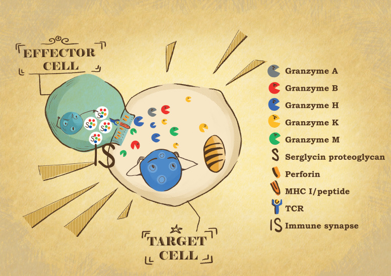
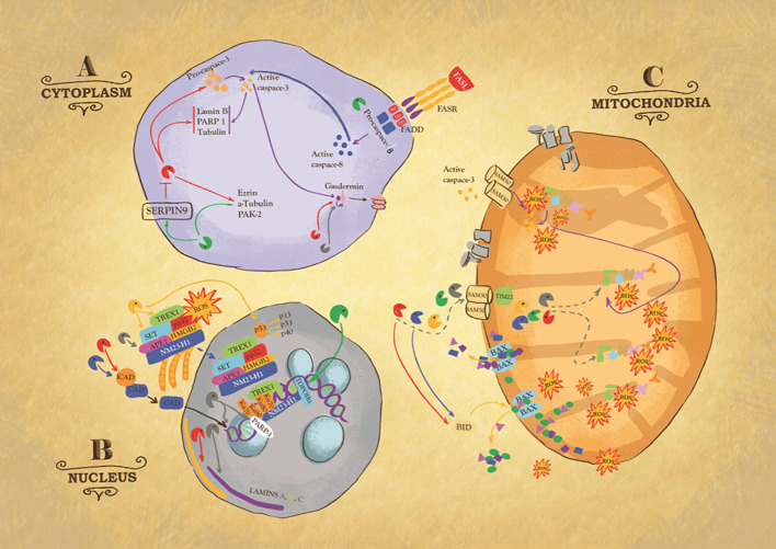
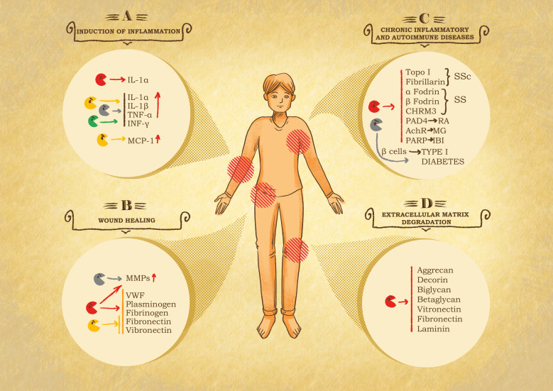
Granzymes are a family of serine proteases, composed of five human members: GA, B, H, M and K. They were first discovered in the 1980s within cytotoxic granules released during NK cell- and T cell-mediated killing. Through their various proteolytic activities, granzymes can trigger different pathways within cells, all of which ultimately lead to the same result, cell death. Over the years, the initial consideration of granzymes as mere cytotoxic mediators has changed due to surprising findings demonstrating their expression in cells other than immune effectors as well as new intracellular and extracellular activities. Additional roles have been identified in the extracellular milieu, following granzyme escape from the immunological synapse or their release by specific cell types. Outside the cell, granzyme activities mediate extracellular matrix alteration via the degradation of matrix proteins or surface receptors. In certain contexts, these processes are essential for tissue homeostasis; in others, excessive matrix degradation and extensive cell death contribute to the onset of chronic diseases, inflammation, and autoimmunity. Here, we provide an overview of both the physiological and pathological roles of granzymes, highlighting their utility while also recognizing how their unregulated presence can trigger the development and/or worsening of diseases.

Keywords: cancer; cytotoxic lymphocytes; granzyme; inflammation; wound healing.


Conflict of interest statement

The authors declare that the research was conducted in the absence of any commercial or financial relationships that could be construed as a potential conflict of interest. The author(s) declared that they were an editorial board member of Frontiers, at the time of submission. This had no impact on the peer review process and the final decision

Figures

Figure 1
Figure 1
Effector: target cell conjugate formation. When the effector cytotoxic T lymphocytes or NK cells recognize their target cell, they form a tight conjugate separated by a narrow space called the immunological synapse (IS). By exocytosis, the effector cells release the content of their cytotoxic granules into the IS. Granzymes are delivered into the target cell by perforin. In the target cells, the granzyme proteases induce the death of the cells through different pathways, affecting different cell compartments.
Figure 2
Figure 2
Intracellular action of granzymes. Action of granzymes in the cytoplasm (A). Granzyme B (GB) can, directly or indirectly via the activation of procaspase 3, cleaves Laminin B, Nuclear Mitotic Apparatus protein (NuMa), Tubulin, Ezrin, p21 activated kinase 2 (PAK-2) and ICAD, inhibitor of the caspase-activated DNase (CAD), to trigger respectively caspase-independent or caspase-dependent cells death. Some of these substrates are also targets of Granzyme M (GM). GM also cleaves GB inhibitor SERPIN-9, suggesting its role in enhancing GB activity or targets FAS-associated death domain protein (FADD), causing the activation of caspase-8. GA, GB, and caspase 3 cleave member of gasdermin (GSDM) family, to trigger pyroptosis, a highly inflammatory type of cell death. Action of granzymes in the nucleus (B). One critical target of GA is endoplasmic reticulum-associated (SET) complex. In presence of ROS, the SET complex is translocated from the endoplasmic reticulum to the nucleus, where its subunit SET, APE1 are cleaved by GA, leading to single stranded DNA nicking. Ku70 and PARP-1 proteins, which activate DNA repair machinery, are also targets of GA. GK also targets SET complex. GK cleaves p53, to produce cleavage fragments with a higher pro-apoptotic activity. GB and GH cleave ICAD, inhibitor of the caspase activated DNase (CAD), ensuring its translocation inside the nucleus where it breaks up DNA. GB and GA target laminin A, B and C disrupting the nuclear lamina. GM cleaves topoisomerase IIa. Action of granzymes in the mitochondria (C). GB, GA, and GM enter mitochondria through the channels SAM50 in the outer membrane and TIM22 in the inner. It is likely that GH and GK use the same mitochondrial entry pathway. Once inside the organelle, GB and GA directly attack NDUFV1, NDUS1 and NDUFS2, subunits of mitochondria NADH: ubiquinone oxidoreductase complex I, causing the production of Reactive Oxygen Species (ROS). Importantly, GB causes mitochondrial outer membrane permeabilization (MOMP) by activating BH3-only protein (BID), which in turn triggers the oligomerization of Bax and/or Bak. Similarly, GH targets BID.
Figure 3
Figure 3
Role of granzymes in pathological settings. (A) Inflammation induction. After leakage from the immune synapse, granzymes can act on the immediate surrounding to potentiate the immune response at the cost of exacerbated inflammation and therefore inflammatory diseases. GB cleaves IL-1α, producing a more active fragment. GA, GK and GM internalization and catalytic activity trigger the secretion of pro-inflammatory cytokines, e.g., IL-1α, IL-1β, TNF-α and INF-γ by immune system cells. GK triggers the production of monocyte chemoattractant protein 1 (MCP-1) by pulmonary fibroblast. Chronic inflammation linked to GB, GA and GK activity has been shown in the onset of rheumatoid arthritis (RA), spinal cord injuries and in chronic obstructive pulmonary disease (COPD). (B) Chronic wound healing. Granzymes can impair or delay provisional matrix formation, angiogenesis and re-epithelization following the cleavage of von Willebrand factor (VWF), plasminogen, fibrinogen, fibronectin and vitronectin and stimulates metalloproteases (MMPs) in fibroblasts, that further exacerbate tissue damage. (C) Systemic autoimmune diseases. Granzymes have been associated with autoimmune disease and cardiovascular, pulmonary, and neurological disorders due to their catalytic activity, resulting in the production or modification of autoantigens. In this context GB is the main actor, cleaving autoantigens involved in scleroderma (SSc) Sjögren syndrome (SS), RA, myasthenia gravis (MG), and ischemic brain injuries (IBI). GA contributes to diabetes type I, due to its participation in pancreatic β-cell degradation. (D) Extracellular matrix degradation. Protein components of the extracellular matrix are target of granzymes, resulting in the restriction of tumor invasion, tissue destruction, neurotoxicity, and autoimmune or inflammatory diseases. GB cleaves proteoglycans such as aggrecan, decorin, biglycan and betaglycan, which compromises the integrity and organization of the matrix. Similarly, GB also digests adhesive proteins such as laminin, fibronectin and vitronectin, essential components in the skin tissue, therefore contributing to skin damage and pathological conditions.

Similar articles

Cited by

References

    1. Lopez-Otin C, Bond JS. Proteases: multifunctional enzymes in life and disease. J Biol Chem. (2008) 283:30433–7. doi: 10.1074/jbc.R800035200 - DOI - PMC - PubMed
    1. van der Velden VH, Hulsmann AR. Peptidases: structure, function and modulation of peptide-mediated effects in the human lung. Clin Exp Allergy. (1999) 29:445–56. doi: 10.1046/j.1365-2222.1999.00462.x - DOI - PMC - PubMed
    1. Lopez-Otin C, Overall CM. Protease degradomics: a new challenge for proteomics. Nat Rev Mol Cell Biol. (2002) 3:509–19. doi: 10.1038/nrm858 - DOI - PubMed
    1. Ciechanover A. Proteolysis: from the lysosome to ubiquitin and the proteasome. Nat Rev Mol Cell Biol. (2005) 6:79–87. doi: 10.1038/nrm1552 - DOI - PubMed
    1. Lee SE, Jeong SK, Lee SH. Protease and protease-activated receptor-2 signaling in the pathogenesis of atopic dermatitis. Yonsei Med J. (2010) 51:808–22. doi: 10.3349/ymj.2010.51.6.808 - DOI - PMC - PubMed

Publication types