. 2024 Apr 8;14(6):2544-2559.
doi: 10.7150/thno.93269. eCollection 2024.

Mechanosensitive protein polycystin-1 promotes periosteal stem/progenitor cells osteochondral differentiation in fracture healing

Affiliations

Mechanosensitive protein polycystin-1 promotes periosteal stem/progenitor cells osteochondral differentiation in fracture healing

Ran Liu et al. Theranostics. .

Abstract

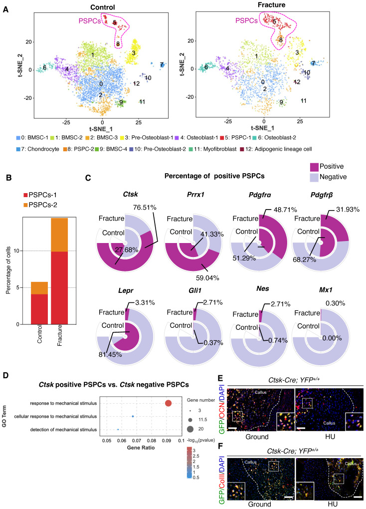
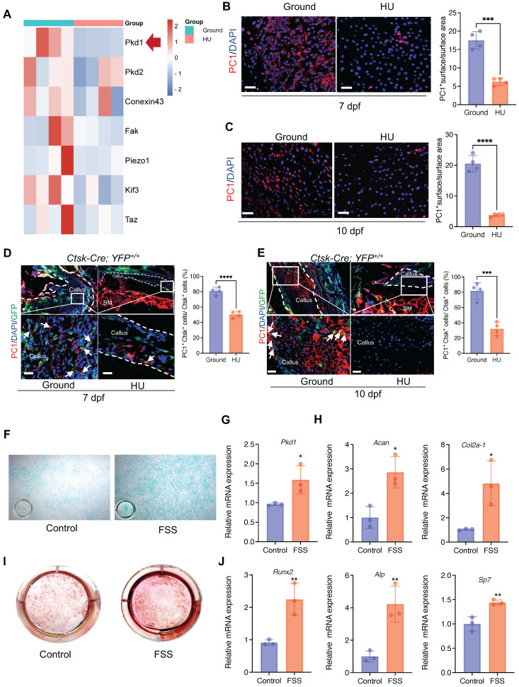
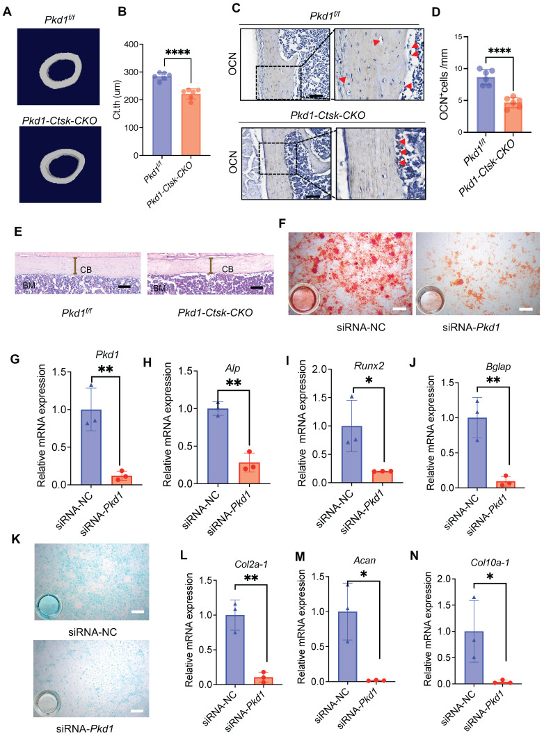
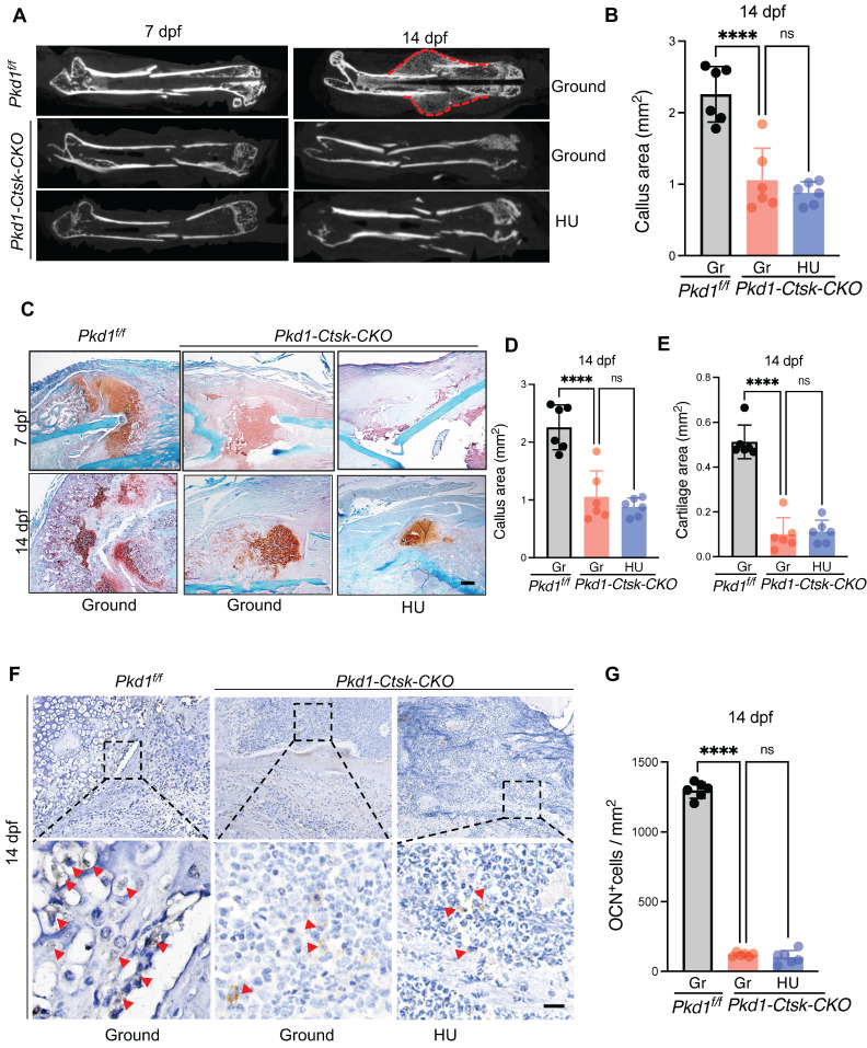
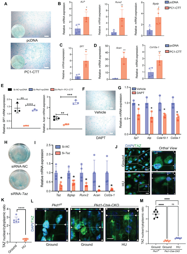
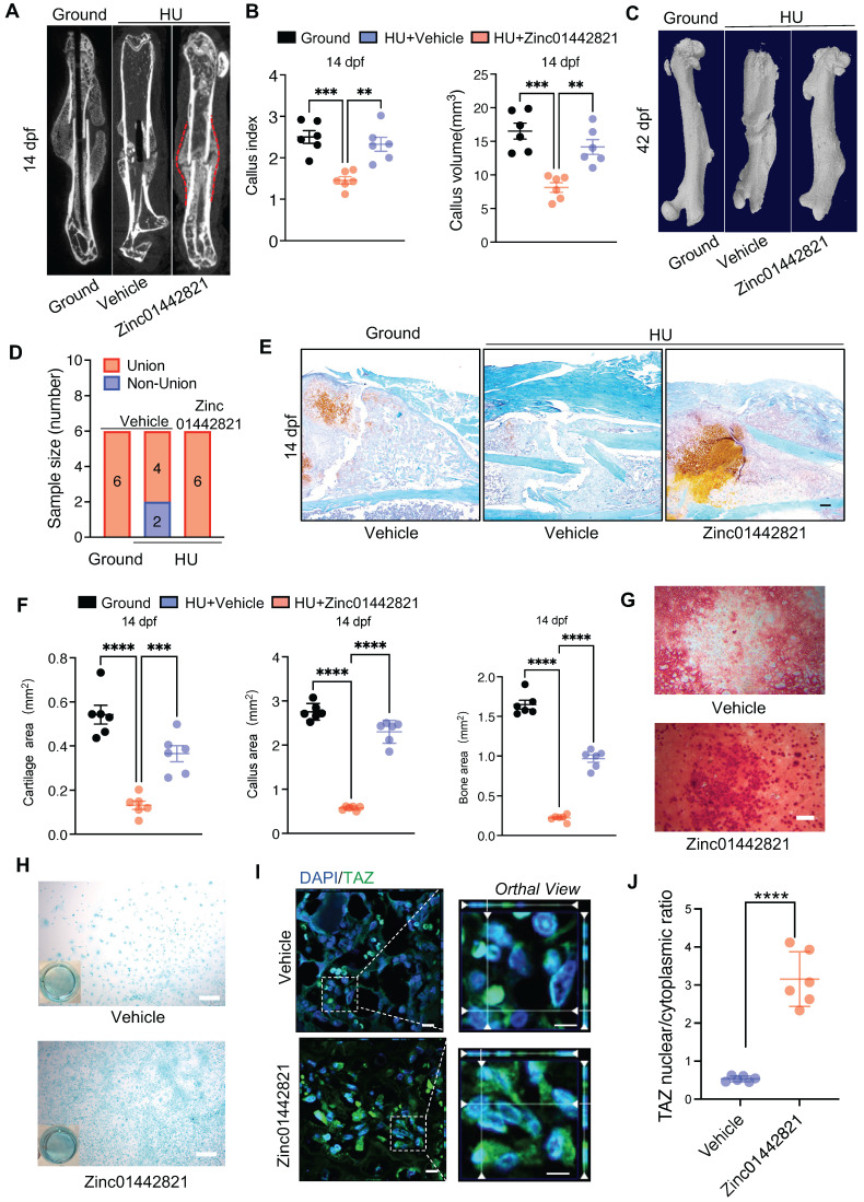
Background: Mechanical forces are indispensable for bone healing, disruption of which is recognized as a contributing cause to nonunion or delayed union. However, the underlying mechanism of mechanical regulation of fracture healing is elusive. Methods: We used the lineage-tracing mouse model, conditional knockout depletion mouse model, hindlimb unloading model and single-cell RNA sequencing to analyze the crucial roles of mechanosensitive protein polycystin-1 (PC1, Pkd1) promotes periosteal stem/progenitor cells (PSPCs) osteochondral differentiation in fracture healing. Results: Our results showed that cathepsin (Ctsk)-positive PSPCs are fracture-responsive and mechanosensitive and can differentiate into osteoblasts and chondrocytes during fracture repair. We found that polycystin-1 declines markedly in PSPCs with mechanical unloading while increasing in response to mechanical stimulus. Mice with conditional depletion of Pkd1 in Ctsk+ PSPCs show impaired osteochondrogenesis, reduced cortical bone formation, delayed fracture healing, and diminished responsiveness to mechanical unloading. Mechanistically, PC1 facilitates nuclear translocation of transcriptional coactivator TAZ via PC1 C-terminal tail cleavage, enhancing osteochondral differentiation potential of PSPCs. Pharmacological intervention of the PC1-TAZ axis and promotion of TAZ nuclear translocation using Zinc01442821 enhances fracture healing and alleviates delayed union or nonunion induced by mechanical unloading. Conclusion: Our study reveals that Ctsk+ PSPCs within the callus can sense mechanical forces through the PC1-TAZ axis, targeting which represents great therapeutic potential for delayed fracture union or nonunion.

Keywords: Fracture healing; Mechanical stress; Periosteal Stem/Progenitor Cells; Polycystin-1.


Conflict of interest statement

Competing Interests: The authors have declared that no competing interest exists.

Figures

Figure 1
Figure 1
Ctsk+ PSPCs are mechanosensitive with osteochondral differentiation potential and contribute to fracture healing. A t-SNE plots showing 13 distinct clusters of cells identified and color-coded from mice fracture models (control group and fracture group). B Stacked bar chart showing the percentages of PSPCs within callus tissue quantified at 7 days post-fracture. C Circular stacked bar plot showing the proportion of positive or negative cells expressing markers gene of PSPCs in control group and fracture group. D GO analysis of differentially expressed genes in Ctsk positive PSPCs or Ctsk negative PSPCs related to mechanical stimuli. E, F Representative IF images of fracture callus at 14 days post-fracture in Ctsk-Cre; YFP+/+ mice, immunostained with OCN (red) or COLLII (red) and GFP (green) antibodies and counterstained with DAPI (blue). White squares indicate magnified areas in callus. Scale bars = 100 μm. Data are presented as means ± SD. Unpaired t test.
Figure 2
Figure 2
Mechanical stimulus affects polycystin-1 level in Ctsk+ PSPCs. A Mechanosensitive gene expression levels in callus from fracture mice at 10 days post-fracture (n = 4). B, C Representative IF images of fracture callus at 7 days and 10 days post-fracture, immunostained with Ctsk (green) and PC1 (red) antibodies and counterstained with DAPI (blue). Dotted squares indicate areas magnified in Periosteum. Scale bars = 100 μm. Immunofluorescence intensity was quantified using ImageJ software (n = 4). D, E Representative IF images of fracture callus at 7- and 10-days post-fracture in Ctsk-Cre; YFP+/+ mice, immunostained with Ctsk (green) and PC1 (red) antibodies and counterstained with DAPI (blue). White squares indicate magnified areas in callus. White arrows indicate co-localization of PC1 level in Ctsk positive cells. Immunofluorescence intensity was quantified using ImageJ software (n = 4). F, I Representative images of Alcian blue staining (F)and Alizarin Red staining (I) of PSPCs with fluid shear stress (FSS). G, H, J qPCR analysis of the efficiency of Pkd1 gene knockout (G), chondrogenic-related genes expression (H) and osteogenic-related genes expression (J) in PSPCs with control and FSS treatment (n = 3). Scale bars = 100 μm. Data are shown as mean ± SD. *p < 0.05, **p < 0.01, ***p < 0.001 and **** p < 0.0001. ns, no significance.
Figure 3
Figure 3
Pkd1 deletion in Ctsk+ PSPCs impairs bone formation. A, B μCT images in femurs from 8-week-old male Pkd1-Ctsk-CKO mice (A) and quantitative analysis of the indicated parameters in Pkd1-Ctsk-CKO mice (B), respectively (n = 6). C, D Representative images of OCN immunohistochemical staining with analysis of number of osteogenic potentials of PSPCs (scalebar = 100 μm; n = 6). E Representative images of H&E staining with analysis of cortical thickness. Scale bars indicates 100 μm. F Representative images of Alizarin Red staining of PSPCs with transfection of siRNA-NC or siRNA-Pkd1. G qPCR analysis of Pkd1 gene expression levels in PSPCs transfected with siRNA-NC or siRNA-Pkd1 (n = 3). Scale bar indicates 100 μm. H-J qPCR analysis of osteogenic-related genes expression in PSPCs transfected with siRNA-NC or siRNA-Pkd1 (n = 3). K Representative images of Alcian blue staining of PSPCs with transfection of siRNA-NC or siRNA-Pkd1. Scale bar indicates 100 μm. L-N qPCR analysis of chondrogenic-related genes expression in PSPCs transfected with siRNA-NC or siRNA-Pkd1 (n = 3). Data are shown as mean ± SD. *p < 0.05, **p < 0.01 and **** p < 0.0001. ns, no significance.
Figure 4
Figure 4
Pkd1 deletion in Ctsk+ PSPCs showed impaired fracture healing and diminished responsiveness to mechanical unloading. A Representative micro-CT images of fractured femurs from Pkd1f/f and Pkd1-Ctsk-CKO mice treated with ground and HU at 7dpf and 14 dpf. B The callus index of fractured femurs from Pkd1f/f and Pkd1-Ctsk-CKO mice treated with ground and HU at 14 dpf. (n = 6). C-E Safranin O staining showed the cartilage callus formation and woven bone area from Pkd1f/f and Pkd1-Ctsk-CKO mice treated with ground and HU at 7 dpf and 14 dpf (n = 6). Scale bar indicates 200 μm. F, G Representative images of OCN immunohistochemical staining with analysis of number of osteoblasts (n = 6). Arrows indicate OCN positive cells. Data are shown as mean ± SD. *p < 0.05, ***p < 0.001 and **** p < 0.0001. ns, no significance.
Figure 5
Figure 5
PC1 regulates osteochondral differentiation potential of PSPCs via its C-terminal tail and downstream TAZ. A Representative image of Alcian blue staining of PSPCs followed by chondrocyte differentiation induction with transfection of control or PC1-CTT plasmids. Scale bar indicates 100 μm. B-D qPCR analysis of osteogenic-related genes expression (B, C) and chondrogenic-related genes expression (D) in PSPCs transfected with control or PC1-CTT plasmids (n = 3-4). E qPCR analysis of Sp7 and Acan in the indicated groups (n = 3). F Alcian blue staining of PSPCs followed by chondrocyte differentiation induction with 10uM DAPT or vehicle treatment. Scale bar indicates 100 μm. G qPCR analysis of Sp7, Alp, Col10 a -1 and Col2a-1 in the 10uM DAPT or vehicle treated groups (n = 3). H Alcian blue staining of PSPCs followed by chondrocyte differentiation induction with siRNA-Taz or siRNA-NC transfected. I qPCR analysis of Taz, Bglap, Runx2, Acan-1 and Col2a-1 of the PSPCs transfected with siRNA-Taz or siRNA-NC (n = 4). J, K Representative immunofluorescence staining images of TAZ (green) in PSPCs isolated from fracture femora in the ground and the HU treated group. Nuclei, DAPI (blue). Dotted squares indicate magnified areas. Scale bar indicates 10 μm. L Representative immunofluorescence staining images of TAZ (green) in PSPCs isolated from Pkd1 fl/fl mice and Pkd1-Ctsk-CKO mice femora. Nuclei, DAPI (blue). Scale bar indicates 5 μm. Data are presented as means ± SD. Unpaired t test. *p < 0.05, **p < 0.01, ***p < 0.001 and **** p < 0.0001.
Figure 6
Figure 6
Zinc01442821 promotes osteochondrogenesis of PSPCs and alleviates unloading-related fracture nonunion. A Representative micro-CT images of fractured femurs from ground and HU mice treated with Vehicle or Zinc01442821 at 14 dpf. B The callus index of fractured femurs from indicated groups at 14 dpf (n = 6). C, D Representative 3D-μCT (C) and quantitative analysis (D) of images of fractured femurs from indicated groups at 42 dpf. E, F Safranin O staining showed the cartilage callus formation from indicated groups at 14 dpf. Scale bar indicates 100 μm. G, H Representative images of Alizarin Red staining (G) and Alcian blue staining (H) of PSPCs treated with vehicle or Zinc01442821. Scale bar indicates 100 μm. I Representative immunofluorescence staining images of TAZ (green) in PSPCs isolated from vehicle or Zinc01442821 mice femora. Nuclei, DAPI (blue). Dotted squares indicate magnified areas. Scale bar indicates 5 μm. J Quantification of the ratio of nuclear TAZ to cytoplasmic TAZ by Welch's t test (n = 6). Data are presented as means ± SD. Unpaired t test, **p < 0.01. **** p < 0.0001.

References

    1. Duda GN, Geissler S, Checa S, Tsitsilonis S, Petersen A, Schmidt-Bleek K. The decisive early phase of bone regeneration. Nat Rev Rheumatol. 2023;19:78–95. - PubMed
    1. Global regional, national burden of bone fractures in 204 countries territories, 1990-2019. a systematic analysis from the Global Burden of Disease Study 2019. Lancet Healthy Longev. 2021;2:e580–e92. - PMC - PubMed
    1. Einhorn TA, Gerstenfeld LC. Fracture healing: mechanisms and interventions. Nat Rev Rheumatol. 2015;11:45–54. - PMC - PubMed
    1. Wildemann B, Ignatius A, Leung F, Taitsman LA, Smith RM, Pesántez R. et al. Non-union bone fractures. Nat Rev Dis Primers. 2021;7:57. - PubMed
    1. Guo Q, Chen N, Patel K, Wan M, Zheng J, Cao X. Unloading-Induced Skeletal Interoception Alters Hypothalamic Signaling to Promote Bone Loss and Fat Metabolism. Adv Sci (Weinh). 2023: e2305042. - PMC - PubMed

Publication types

Substances