Review
. 2024 Aug 1;327(2):C278-C290.
doi: 10.1152/ajpcell.00613.2023. Epub 2024 Apr 22.

Involvement of kinesins in skeletal dysplasia: a review

Affiliations
Review

Involvement of kinesins in skeletal dysplasia: a review

Roufaida Bouchenafa et al. Am J Physiol Cell Physiol. .

Abstract

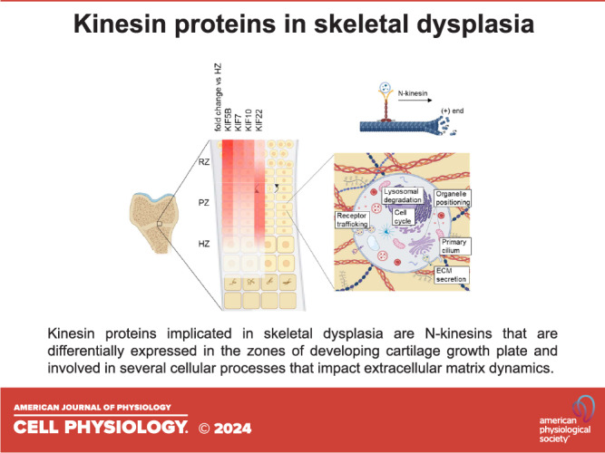
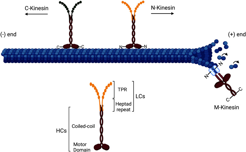
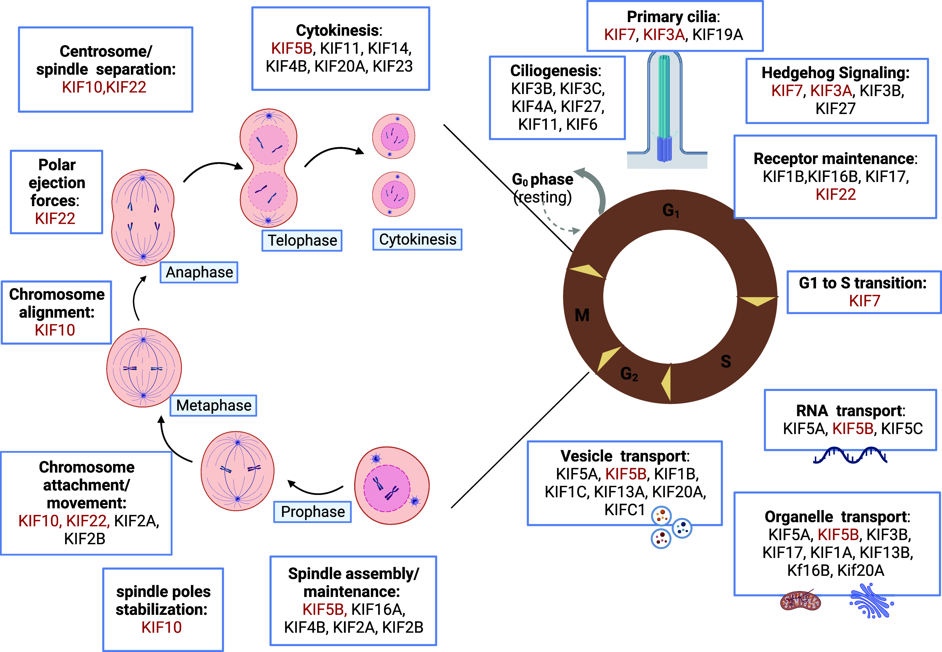
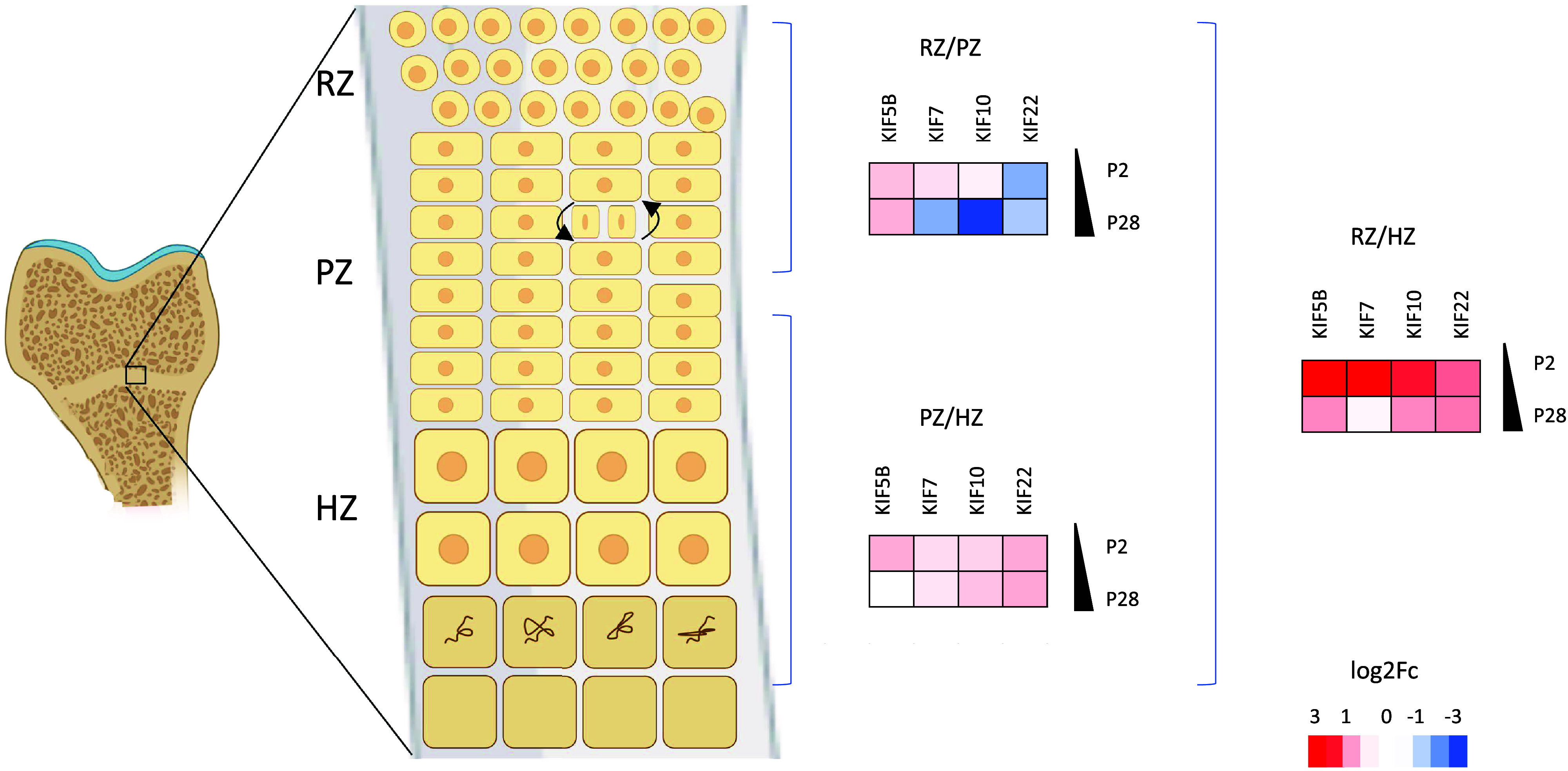
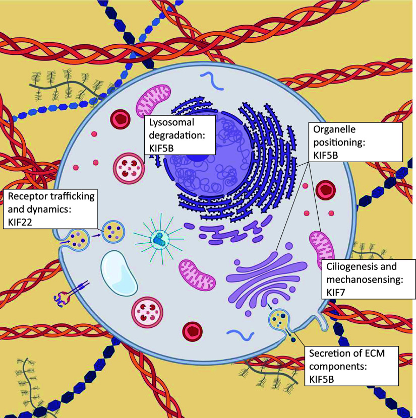
Skeletal dysplasias are group of rare genetic diseases resulting from mutations in genes encoding structural proteins of the cartilage extracellular matrix (ECM), signaling molecules, transcription factors, epigenetic modifiers, and several intracellular proteins. Cell division, organelle maintenance, and intracellular transport are all orchestrated by the cytoskeleton-associated proteins, and intracellular processes affected through microtubule-associated movement are important for the function of skeletal cells. Among microtubule-associated motor proteins, kinesins in particular have been shown to play a key role in cell cycle dynamics, including chromosome segregation, mitotic spindle formation, and ciliogenesis, in addition to cargo trafficking, receptor recycling, and endocytosis. Recent studies highlight the fundamental role of kinesins in embryonic development and morphogenesis and have shown that mutations in kinesin genes lead to several skeletal dysplasias. However, many questions concerning the specific functions of kinesins and their adaptor molecules as well as specific molecular mechanisms in which the kinesin proteins are involved during skeletal development remain unanswered. Here we present a review of the skeletal dysplasias resulting from defects in kinesins and discuss the involvement of kinesin proteins in the molecular mechanisms that are active during skeletal development.

Keywords: chondrocyte; kinesin; microtubules; motor proteins; skeletal dysplasia.


Conflict of interest statement

No conflicts of interest, financial or otherwise, are declared by the authors.

Figures

None
Graphical abstract
Figure 1.
Figure 1.
Molecular structure of kinesins. Kinesin dimers comprise two heavy chains (HCs) with a microtubule-binding site (motor domain) and a coiled-coil domain, and two light chains (LCs) comprising the tetratricopeptide repeat domain (KLCTPR) and a heptad repeat that associate to the coiled-coil domain of HCs (80). The position of the motor domain defines the kinesin classes: N-Kinesins have an N-terminus motor domain and drive plus-ended microtubule movement, M-Kinesins have their motor domain in the middle and are MT depolymerizers, and C-kinesin have a carboxy (C)-terminal motor domain and mediate minus-end-directed movement (81, 82). Created with BioRender.com.
Figure 2.
Figure 2.
Kinesins play a role in multiple cell processes. Kinesins ensure specific functions during different phases of cell division, mediating chromosome movement and alignment, spindle formation, and regulating microtubule-kinetochore interaction. They are also involved in the maintenance of the primary cilia structure and function, and drive intracellular transport of organelles, vesicles, receptors, and RNA along microtubules. The kinesins implicated in skeletal dysplasia have been highlighted in red. Created with BioRender.com.
Figure 3.
Figure 3.
KIF5B, KIF7, KIF10, and KIF22 are differentially expressed in the zones of murine epiphyseal cartilage during development. Fold change data were obtained from SkeletalVis (106), and heat maps showing differential expression (log2FC) were generated. Key: HZ, hypertrophic zone; PZ, proliferative zone; P2, postnatal day 2; P28, postnatal day 28; RZ, resting zone. Created with BioRender.com.
Figure 4.
Figure 4.
Kinesin motor proteins mediate ECM-related processes. Kinesin molecules implicated in skeletal dysplasia play roles in organelle positioning (including the endoplasmic reticulum and Golgi apparatus) and receptor recycling, and contribute to the intracellular transport of vesicles containing ECM components. Kinesins also play roles in ciliogenesis and mechanosensing. ECM, extracellular matrix. Created with BioRender.com.

Similar articles

Cited by

References

    1. Warman ML, Cormier‐Daire V, Hall C, Krakow D, Lachman R, Lemerrer M, Mortier G, Mundlos S, Nishimura G, Rimoin DL, Robertson S, Savarirayan R, Sillence D, Spranger J, Unger S, Zabel B, Superti‐Furga A. Nosology and classification of genetic skeletal disorders: 2010 revision. Am J Med Genet A 155A: 943–968, 2011. doi:10.1002/ajmg.a.33909. - DOI - PMC - PubMed
    1. Krakow D, Rimoin DL. The skeletal dysplasias. Genet Med 12: 327–341, 2010. doi:10.1097/GIM.0b013e3181daae9b. - DOI - PubMed
    1. Orioli IM, Castilla EE, Barbosa-Neto JG. The birth prevalence rates for the skeletal dysplasias. J Med Genet 23: 328–332, 1986. doi:10.1136/jmg.23.4.328. - DOI - PMC - PubMed
    1. Kolambage YD, Walpita YN, Liyanage UA, Dayaratne BMKDR, Dissanayake VHW. The burden of hospital admissions for skeletal dysplasias in Sri Lanka: a population-based study. Orphanet J Rare Dis 18: 279, 2023. doi:10.1186/s13023-023-02884-2. - DOI - PMC - PubMed
    1. Unger S, Ferreira CR, Mortier GR, Ali H, Bertola DR, Calder A, Cohn DH, Cormier-Daire V, Girisha KM, Hall C, Krakow D, Makitie O, Mundlos S, Nishimura G, Robertson SP, Savarirayan R, Sillence D, Simon M, Sutton VR, Warman ML, Superti-Furga A. Nosology of genetic skeletal disorders: 2023 revision. Am J Med Genet A 191: 1164–1209, 2023. doi:10.1002/ajmg.a.63132. - DOI - PMC - PubMed

LinkOut - more resources