Review
. 2024 Aug 30;73(S1):S401-S412.
doi: 10.33549/physiolres.935306. Epub 2024 Apr 22.

Look for the Scaffold: Multifaceted Regulation of Enzyme Activity by 14-3-3 Proteins

Affiliations
Review

Look for the Scaffold: Multifaceted Regulation of Enzyme Activity by 14-3-3 Proteins

V Obsilova et al. Physiol Res. .

Abstract

Enzyme activity is regulated by several mechanisms, including phosphorylation. Phosphorylation is a key signal transduction process in all eukaryotic cells and is thus crucial for virtually all cellular processes. In addition to its direct effect on protein structure, phosphorylation also affects protein-protein interactions, such as binding to scaffolding 14-3-3 proteins, which selectively recognize phosphorylated motifs. These interactions then modulate the catalytic activity, cellular localisation and interactions of phosphorylated enzymes through different mechanisms. The aim of this mini-review is to highlight several examples of 14-3-3 protein-dependent mechanisms of enzyme regulation previously studied in our laboratory over the past decade. More specifically, we address here the regulation of the human enzymes ubiquitin ligase Nedd4-2, procaspase-2, calcium-calmodulin dependent kinases CaMKK1/2, and death-associated protein kinase 2 (DAPK2) and yeast neutral trehalase Nth1.


Conflict of interest statement

Conflict of Interest: There is no conflict of interest.

Figures

Fig. 1
Fig. 1
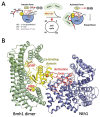
Allosteric regulation of Nth1 by 14-3-3 protein. (A) Schematic model of 14-3-3-dependent regulation of Nth1. The inactive form of Nth1 is unable to hydrolyze trehalose to glucose. The N-terminal segment of Nth1 contains five phosphorylation sites, of which four are recognized by PKA and the other by Cdk1. The Ser83-containing motif serves as a gatekeeper, the dominant site for 14-3-3 protein binding, while the Ser60-containing motif functions as a secondary site [36,38,40]. An alternative secondary site is the Ser66-containing motif phosphorylated by Cdk1. The Ser20 and Ser21 sites are part of the binding site for the phosphatase, but this interaction is suppressed by phosphorylation [44]. Phosphorylated Nth1 is recognized by the yeast 14-3-3 protein during the G1 to S cell cycle transition [43]. 14-3-3 protein binding induces a conformational change in both the calcium and catalytic do-mains, creating a loop with all the residues necessary for catalysis. The active form of Nth1 then begins to metabolize trehalose to glucose. (B) Crystal structure of the Bmh1:Nth1 complex (PDB: 5N6N) [40]. The protomers of the Bmh1 homodimer are shown in two shades of green. The N-terminal extension and the calcium-binding domain are shown in yellow, and the catalytic domain is highlighted in blue. The phosphorylated Ser60 and Ser83, the regulatory loop and its two residues crucial for catalysis (Glu690 and Tyr691) are shown in red. The calcium ion is shown in orange. The figure was prepared with PyMOL (https://pymol.org/2/).
Fig. 2
Fig. 2
Ribbon representation of SAXS-based structural models of select 14-3-3 protein complexes. The protomers of the 14-3-3γ and 14-3-3η homodimer are shown in two shades of green in all panels. (A) The best-scoring CORAL model of the Nedd4-2:14-3-3η complex constructed using crystal structures of HECT domain (PDB ID: 5HPK) [96], solution structures of WW1-3 domains (PDB ID: 1WR3, 1WR4, 1WR7) and 14-3-3γ with bound Nedd4-2 phosphopeptides pSer342 and pSer448 (PDB ID 6ZBT, 6ZC9)[56]. WW2-4 are shown in yellow, orange and sand, the N- and C-lobes of the HECT domain are shown in blue and magenta, respectively. (B) The best-scoring AllosMod-FoXS model of the procaspase-2:14-3-3γ complex [73] constructed using the crystal structure of caspase-2 (PDB ID: 3R7S) [97] and the 14-3-3γ with bound caspase-2 peptide phosphorylated on Ser139 and Ser164 (PDB ID 6SAD) [74]. p19 and p12 domains of procaspase-2 are shown in blue and magenta, respectively. (C) The best-scoring CORAL model of the DAPK2:14-3-3γ complex constructed using crystal structures of autoinhibited DAPK2 (PDB ID: 2A2A) [80,81] and 14-3-3γ with bound C-terminal DAPK2 phophopeptidepeptide pThr369 (PDB ID: 7A6R) [83]. The unstructured segments missing in the crystal structures were modelled as dummy residue chains, shown as spheres. Two protomers of DAPK2 dimer are shown in blue and magenta. (D) The best-scoring AllosMod-FoXS model of the pCaMKK1:14-3-3γ complex constructed using crystal structures of the kinase domain of CaMKK1 (PDB ID: 6CD6) and 14-3-3γ with bound CaMKK2 phosphopeptides (PDB ID: 6FEL and 6EWW) [94,95]. The position of the active site is indicated by the position of inhibitor in the crystal structure of CaMKK1 (yellow sticks). The figure was prepared with PyMOL (https://pymol.org/2/).

Similar articles

References

    1. Choi KY, Satterberg B, Lyons DM, Elion EA. Ste5 tethers multiple protein kinases in the MAP kinase cascade required for mating in S. cerevisiae. Cell. 1994;78:499–512. doi: 10.1016/0092-8674(94)90427-8. - DOI - PubMed
    1. Rubin CS, Rosen OM. Protein phosphorylation. Annu Rev Biochem. 1975;44:831–887. doi: 10.1146/annurev.bi.44.070175.004151. - DOI - PubMed
    1. Reinhardt HC, Yaffe MB. Phospho-Ser/Thr-binding domains: navigating the cell cycle and DNA damage response. Nat Rev Mol Cell Biol. 2013;14:563–580. doi: 10.1038/nrm3640. - DOI - PubMed
    1. Furukawa Y, Ikuta N, Omata S, Yamauchi T, Isobe T, Ichimura T. Demonstration of the phosphorylation-dependent interaction of tryptophan hydroxylase with the 14-3-3 protein. Biochem Biophys Res Commun. 1993;194:144–149. doi: 10.1006/bbrc.1993.1796. - DOI - PubMed
    1. Fantl WJ, Muslin AJ, Kikuchi A, Martin JA, MacNicol AM, Gross RW, Williams LT. Activation of Raf-1 by 14-3-3 proteins. Nature. 1994;371:612–614. doi: 10.1038/371612a0. - DOI - PubMed

Substances